The Family Of Fatty Skin Deposits Explained: Every Type Of Xanthoma, What Each One Says About Your Blood Fats, How They’re Diagnosed, And Where Eyelid Xanthelasma Fits
A xanthoma is a yellowish deposit of fat-filled cells in the skin, tendons or, occasionally, internal organs. There are several types, from the common, soft yellow spots on the eyelids called xanthelasma to firm tendon lumps, crops of eruptive bumps and creased palms, and each points to different blood-fat problems. Most are harmless in themselves, but some are strong signs of an inherited or serious lipid disorder, so every new xanthoma deserves a doctor’s check.
A xanthoma is a benign, yellowish lump or patch made of cholesterol-filled foam cells. Types include xanthelasma on the eyelids, tendon, eruptive, tuberous, palmar and plane xanthomas. Tendon and eruptive types strongly suggest a lipid disorder; about half of people with xanthelasma have one. See a doctor for blood tests.
Key Takeaways
- A xanthoma is a deposit of cholesterol-filled foam cells, usually in skin or tendons.
- Xanthelasma on the eyelids is the type people most often notice.
- Tendon xanthomas are a hallmark of inherited high cholesterol.
- Eruptive xanthomas appear with triglycerides above about 1,000 mg/dL.
- Some types fade when the blood-fat problem is managed; eyelid plaques rarely do.
- Every new xanthoma is worth a blood test.
On This Page 17 sections
What Is A Xanthoma?
A xanthoma is a yellow, orange or skin-colored deposit made of immune cells packed with fat. The name comes from the Greek for “yellow” and “growth”. Xanthomas are benign, but they often reflect how your body handles cholesterol and triglycerides.
Xanthomas vary in size from a few millimeters to several centimeters and can be flat, soft, firm or bumpy depending on the type. They’re found most often on the eyelids, over tendons, on the elbows, knees, buttocks and hands, and in the creases of the palms. The plural is xanthomas or xanthomata, and when many appear together doctors may call it xanthomatosis.
Our guides to xanthomas, the xanthoma definition and xanthomata explain the terms in more depth.

How Do Xanthomas Form?
Fat-carrying particles leak from small blood vessels into the tissue, where immune cells called macrophages swallow them. Overloaded with fat, these cells become foam cells and cluster together to form a xanthoma.
StatPearls describes this process for eyelid xanthelasma: lipid-laden macrophages become foam cells in the deeper layer of the skin around small vessels, with some inflammation, helped by blinking and vessels that become leakier with age.[1] The same basic process happens elsewhere; what differs is which fats are high, how high they are, and where the tissue is under stress, such as tendons, elbows and knees.
That’s why the type and location of a xanthoma can hint at which blood-fat problem is behind it. Our page on what a xanthoma is covers the biology in more detail.

What Are The Main Types Of Xanthoma?
Xanthelasma (eyelids), tendon xanthomas, eruptive xanthomas, tuberous xanthomas, palmar xanthomas and plane xanthomas, plus rare forms such as xanthoma disseminatum and xanthomas inside the body.
- Xanthelasma. Soft yellow plaques on the eyelids.
- Tendon. Firm, smooth lumps over the Achilles, knuckles, elbows or knees.
- Eruptive. Sudden crops of small yellow-red bumps, often on the buttocks, arms and legs.
- Tuberous. Firm, rounded nodules over the elbows and knees.
- Palmar. Yellow streaks in the creases of the palms and fingers.
- Plane. Flat yellow patches, sometimes widespread.
Our pages on what xanthomas are and skin xanthomas describe how each looks.

Is Xanthelasma A Type Of Xanthoma?
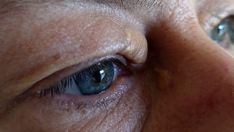
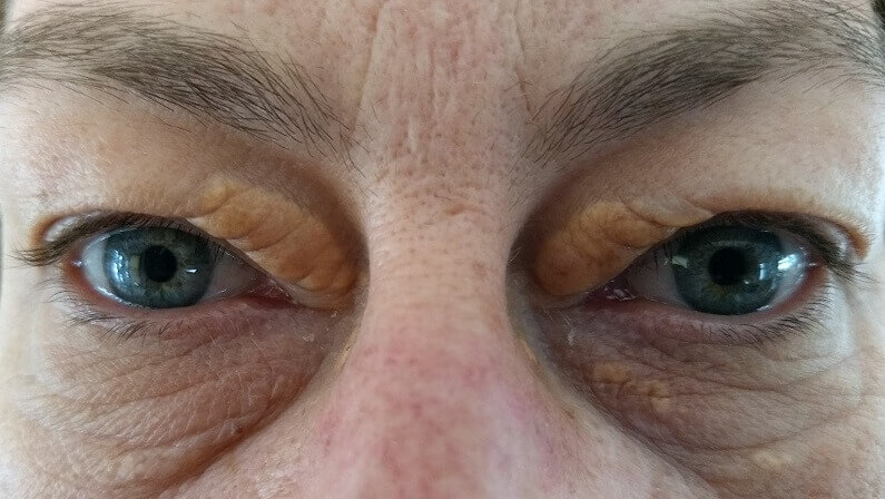
Yes. Xanthelasma is the eyelid type of xanthoma and the one people most often notice. It affects about 1.1% of women and 0.3% of men, and about half of people with it have a lipid disorder.
Xanthelasma palpebrarum appears as soft, flat or slightly raised yellow plaques, usually near the inner corners of the upper or lower lids, most often between 35 and 55.[1] Unlike tendon or eruptive xanthomas, it often appears with normal cholesterol, but about 50% of people with it have a primary lipid disorder or a secondary cause such as diabetes, an underactive thyroid, a kidney or liver condition, or retinoid or estrogen medicines.[1]
Eyelid plaques rarely fade, even when cholesterol improves, so many people choose to remove them. Our guides to xanthelasma, what xanthelasma is and the eyelid xanthoma go further.

If your xanthoma is eyelid xanthelasma, your doctor has checked the cause, and you’d like to remove the plaques at home, this is the cream we make for it.
What Are Tendon Xanthomas?

Firm, smooth, skin-colored lumps over tendons, most often the Achilles, the backs of the hands, elbows and knees. They’re a hallmark of familial hypercholesterolemia and, more rarely, of a condition called cerebrotendinous xanthomatosis.
Familial hypercholesterolemia affects about 1 in 250 to 1 in 500 people, passes to each child with a 50% chance, and causes very high LDL from birth. Tendon xanthomas on the Achilles, elbows, hands, knees or feet, and xanthelasma, are typical signs. Without management, about 50% of men have a coronary event by 50 and about 30% of women by 60; statins are the first-line medicine.[2]
Cerebrotendinous xanthomatosis is a rare inherited condition caused by changes in the CYP27A1 gene. It typically causes diarrhea in infancy, cataracts in childhood in about 75% of people, tendon xanthomas in the teens or twenties and nerve problems in adulthood; chenodeoxycholic acid is the standard medicine, and early care appears to prevent symptoms.[3] Our pages on tendon xanthomas and Achilles tendon xanthoma explain more.

What Are Eruptive Xanthomas?
Sudden crops of small, yellow-red bumps, often itchy or tender, usually on the buttocks, shoulders, arms and legs. They appear when triglycerides are very high, typically above 1,000 mg/dL, and need prompt medical attention.
StatPearls links eruptive xanthomas with triglycerides above 1,000 mg/dL, notes that uncontrolled diabetes raises triglycerides, and warns that such high levels carry a risk of pancreatitis.[4] Very high triglycerides can even turn the blood vessels at the back of the eye a milky color, a sign called lipemia retinalis.[4]
The good news is that eruptive xanthomas usually settle once triglycerides are brought down. Our guide to eruptive xanthoma covers the causes and next steps.

What Are Tuberous And Palmar Xanthomas?
Tuberous xanthomas are firm, yellow, rounded nodules, usually over the elbows and knees. Palmar xanthomas are yellow streaks in the creases of the palms and fingers. Both are classic signs of dysbetalipoproteinemia.
Dysbetalipoproteinemia, also called type III hyperlipoproteinemia, raises both cholesterol and triglycerides. StatPearls lists palmar and tuberous xanthomas as its typical skin signs and notes that palmar xanthomas fade once the condition is managed.[5] Tuberous xanthomas can also appear in familial hypercholesterolemia.
Our pages on tuberous xanthoma and palmar xanthoma go into each.

What Are Plane Xanthomas?
Flat, yellow-orange patches in the skin. They can appear in skin folds, on the neck and trunk, or more widely. When they’re widespread with normal blood fats, doctors check for a blood protein disorder.
Diffuse plane xanthoma with normal lipid levels has been linked with monoclonal gammopathy, an abnormal protein made by one group of blood cells, which can occur on its own or with conditions such as myeloma.[6] That’s why widespread flat xanthomas, especially with normal cholesterol, need a thorough medical work-up. Plane xanthomas in the palm creases overlap with the palmar type above. Our page on plane xanthoma explains more.

What Are The Rare Types Of Xanthoma?
Xanthoma disseminatum, a rare condition with many small yellow-brown bumps in skin folds and sometimes the airways, and xanthomas inside the body, such as gastric xanthoma in the stomach lining.
Xanthoma disseminatum is uncommon and usually occurs with normal blood fats, so it’s thought to arise from a different process than the lipid-driven types. Gastric xanthoma is a small yellow-white patch seen during endoscopy of the stomach; despite the shared name, it’s a separate condition with its own significance. Our pages on xanthoma disseminatum, gastric xanthoma and xanthomatosis cover these in detail.

Can Children And Young People Get Xanthomas?
Yes, though it’s uncommon, and it’s always worth investigating. Xanthomas in a child or young adult usually point to an inherited condition such as familial hypercholesterolemia or, rarely, cerebrotendinous xanthomatosis.
In familial hypercholesterolemia, LDL is high from birth, and in the more severe forms tendon or skin xanthomas can appear in childhood.[2] In cerebrotendinous xanthomatosis, childhood cataracts and long-lasting diarrhea in infancy often come years before the tendon lumps, which typically show in the teens or twenties.[3] StatPearls also advises considering an inherited lipid disorder whenever eyelid xanthelasma appears at a young age.[1]
Because both conditions respond far better to early care, and because family members may share them, a young person with any type of xanthoma should see a doctor, and relatives may be offered testing too.

Types Of Xanthoma Compared
| Type | Where | Looks Like | Often Linked With |
|---|---|---|---|
| Xanthelasma | Eyelids | Soft, flat or slightly raised yellow plaques | A lipid disorder in about half; often normal cholesterol |
| Tendon | Achilles, hands, elbows, knees | Firm, smooth lumps | Familial hypercholesterolemia; rarely CTX |
| Eruptive | Buttocks, shoulders, arms, legs | Crops of small yellow-red bumps | Triglycerides above about 1,000 mg/dL, diabetes |
| Tuberous | Elbows, knees | Firm, rounded yellow nodules | Dysbetalipoproteinemia, familial hypercholesterolemia |
| Palmar | Palm and finger creases | Yellow streaks | Dysbetalipoproteinemia |
| Plane | Skin folds, neck, trunk | Flat yellow-orange patches | Lipid disorders; if widespread with normal lipids, monoclonal gammopathy |
| Disseminatum | Skin folds, sometimes airways | Many small yellow-brown bumps | Usually normal blood fats |
What Can A Xanthoma Say About Your Health?

It depends on the type. Tendon, eruptive, tuberous and palmar xanthomas strongly suggest a specific lipid disorder. Xanthelasma is a weaker signal, linked with a lipid disorder in about half of cases. Widespread plane xanthomas with normal fats can point to a blood protein disorder.
- Very high LDL from birth. Tendon xanthomas and early xanthelasma suggest familial hypercholesterolemia.[2]
- Very high triglycerides. Eruptive xanthomas, often with uncontrolled diabetes.[4]
- Both raised. Palmar and tuberous xanthomas suggest dysbetalipoproteinemia.[5]
- Normal lipids, widespread flat patches. Check for monoclonal gammopathy.[6]
- Eyelid plaques. A lipid disorder in about half; secondary causes include diabetes, thyroid, kidney and liver conditions.[1]
Our page on whether xanthelasma indicates raised cholesterol explains the eyelid link.

How Are Xanthomas Diagnosed?
Usually by looking at them, followed by blood tests to find the cause. A small biopsy is taken if the diagnosis is unclear, and some types need further tests.
- Examination. The type, location and number of xanthomas give strong clues.
- Lipid profile. A fasting test of total cholesterol, LDL, HDL and triglycerides.[1]
- Other blood tests. Liver panel, thyroid tests and fasting glucose, with HbA1c in diabetes.[1]
- Specialist tests if needed. Genetic testing for inherited disorders, or protein tests for widespread plane xanthomas.
- Biopsy if unsure. A small sample confirms foam cells under the microscope.[1]

Can Xanthomas Be Managed Or Removed?
Yes. The first step is managing the blood-fat problem or other cause, which can make eruptive and palmar xanthomas fade. Xanthelasma and some tendon or tuberous xanthomas usually persist and may be removed.
Managing the cause can include diet, activity, cholesterol- or triglyceride-lowering medicine, diabetes and thyroid care, or specific medicines such as chenodeoxycholic acid for cerebrotendinous xanthomatosis.[2][3] For eyelid xanthelasma, removal options include an at-home cream made for xanthelasma removal, laser and surgery; recurrence is generally under 50%.[1] Larger body xanthomas are sometimes removed surgically by a specialist.
Our pages on xanthoma removal and clinic options and xanthelasma home removal cover the choices.

Which Doctor Should You See About A Xanthoma?
Start with your family doctor for the blood tests. Depending on the type, you may be referred to a lipid specialist, endocrinologist, dermatologist, eye or eyelid specialist, or hematologist.
Seek help promptly if xanthomas appear suddenly in crops, if you have tendon lumps, if you’re young, or if close relatives had early heart disease, since these point to a significant lipid disorder.[2][4] Severe stomach pain with eruptive xanthomas needs urgent care because of the pancreatitis risk.[4]

What’s The Bottom Line On Xanthoma?
A xanthoma is a benign deposit of cholesterol-filled cells. Xanthelasma on the eyelids is the type most people see; tendon, eruptive, tuberous, palmar and plane xanthomas are less common and often point to a specific lipid or blood disorder. Every new xanthoma deserves a blood test, managing the cause comes first, and eyelid plaques can be removed if you’d like them gone.
Xanthel® is a cosmetic cream made for xanthelasma removal at home, sold by Xanthelasma.com, for the eyelid type only. You’ll find it in the xanthelasma section above and at the foot of this page, or read more about Xanthel® xanthelasma removal cream. For everything else, browse all our xanthelasma and xanthoma guides.
Common Questions About Xanthoma
What Is A Xanthoma?
A benign yellowish deposit of fat-filled immune cells, called foam cells, in the skin, tendons or occasionally internal organs.
What Are The Different Types Of Xanthoma?
Xanthelasma on the eyelids, tendon, eruptive, tuberous, palmar and plane xanthomas, plus rare forms such as xanthoma disseminatum.
Is Xanthelasma A Type Of Xanthoma?
Yes, xanthelasma is the eyelid type of xanthoma and the one people most often notice.
Are Xanthomas Dangerous?
The deposits are benign, but some types signal serious lipid or blood disorders that raise heart or pancreatitis risk, so they should be checked.
Do Xanthomas Mean I Have High Cholesterol?
Often but not always; tendon and eruptive types strongly suggest a lipid disorder, while about half of people with xanthelasma have normal levels.
Do Xanthomas Go Away?
Eruptive and palmar xanthomas often fade once the blood-fat problem is managed, but xanthelasma and many tendon xanthomas usually persist.
Can Xanthomas Be Removed?
Yes; eyelid xanthelasma can be removed with a cream made for it, laser or surgery, and larger body xanthomas are sometimes removed surgically.
Which Doctor Should I See About A Xanthoma?
Start with your family doctor for blood tests, who may refer you to a lipid specialist, dermatologist, eyelid specialist or hematologist.
Sources
- Blair K. Xanthelasma Palpebrarum. StatPearls, National Library of Medicine (NCBI Bookshelf), updated 2026.
- Ison HE, Clarke SL, Knowles JW. Familial Hypercholesterolemia. GeneReviews, National Library of Medicine (NCBI Bookshelf), revised 2025.
- Federico A, Gallus GN. Cerebrotendinous Xanthomatosis. GeneReviews, National Library of Medicine (NCBI Bookshelf), revised 2024.
- Mishra C, Tripathy K. Lipemia Retinalis. StatPearls, National Library of Medicine (NCBI Bookshelf), updated 2026.
- Javvaji A, Can AS, Sharma S. Dysbetalipoproteinemia. StatPearls, National Library of Medicine (NCBI Bookshelf), updated 2024.
- Szalat R, et al. Pathogenesis and treatment of xanthomatosis associated with monoclonal gammopathy. Blood, 2011.