When A Xanthoma Appears On The Eyelid It’s Usually Xanthelasma: How It Differs From Other Xanthomas, The Rare Exceptions, And Removal
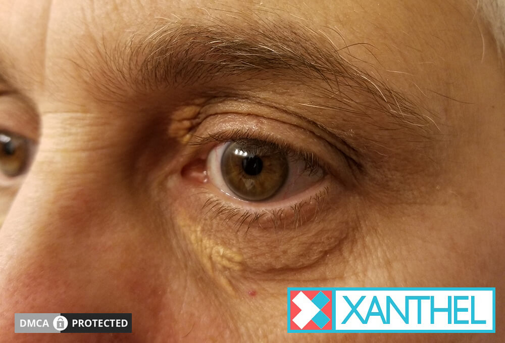
A xanthoma on the eyelid is a soft, yellow deposit of fat-filled cells in the eyelid skin, and doctors almost always call it xanthelasma. It’s the most common type of xanthoma and usually harmless, but it can point to a cholesterol problem, and a few rarer eyelid conditions can look similar. Here’s how an eyelid xanthoma fits into the xanthoma family, what it can mean, and how to remove it.
An eyelid xanthoma is xanthelasma: a soft yellow plaque of fat-filled immune cells, usually near the inner corners of the eyelids. It’s the most common xanthoma and is often harmless, but about half of people have a blood fat disorder. Rarely, firm or ulcerating yellow eyelid lesions are something else, so a doctor should confirm it once.
Key Takeaways
- A xanthoma on the eyelid is called xanthelasma.
- It’s the most common type of xanthoma.
- About half of people with it have a blood fat disorder.
- Eyelid xanthomas are usually simpler than xanthomas elsewhere on the body.
- Rare conditions can cause firmer yellow eyelid lesions that need a doctor.
- Eyelid xanthomas can be removed at a clinic or at home.
On This Page 15 sections
What Is A Xanthoma On The Eyelid?
It’s a soft, yellow, flat or slightly raised deposit of fat-filled immune cells in the eyelid skin. When it’s on the eyelids, doctors call it xanthelasma, or xanthelasma palpebrarum.
Xanthomas are deposits made of foam cells: immune cells called macrophages that have filled up with fat. They can form in the skin, tendons and other tissues. On the eyelids, they gather around tiny blood vessels in the deeper layer of the skin, with some inflammation, and show through the thin skin as a yellow patch.[1]
Eyelid xanthomas affect around 1.1% of women and 0.3% of men, most often between 35 and 55, and they’re the most common type of xanthoma.[1] Our guides to xanthelasma, xanthelasma palpebrarum and eyelid xanthomas cover the eyelid type in depth.

Is An Eyelid Xanthoma The Same As Xanthelasma?
Yes. “Xanthoma” is the umbrella term for any fat-filled deposit, and “xanthelasma” is the specific name for one on the eyelids. An eyelid xanthoma and xanthelasma are the same thing.
Xanthoma
- The family name for fat-filled deposits
- Can form on the eyelids, tendons, skin and elsewhere
- Plural: xanthomas or xanthomata
- Many types point to a blood fat disorder
Xanthelasma
- The name for a xanthoma on the eyelids
- The most common type of xanthoma
- Full name: xanthelasma palpebrarum
- Often a cosmetic concern
You may also see eyelid xanthomas called cholesterol deposits on the eyelids or yellow spots on the eyelids. Our guides to the xanthoma definition, what a xanthoma is and xanthomas explain the wider family.

What Does An Eyelid Xanthoma Look Like?
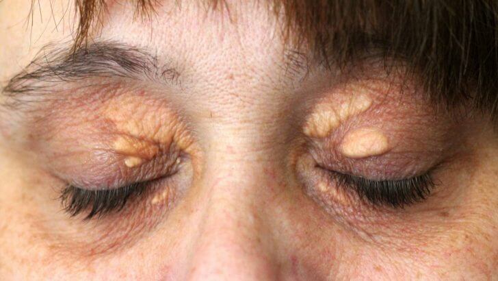
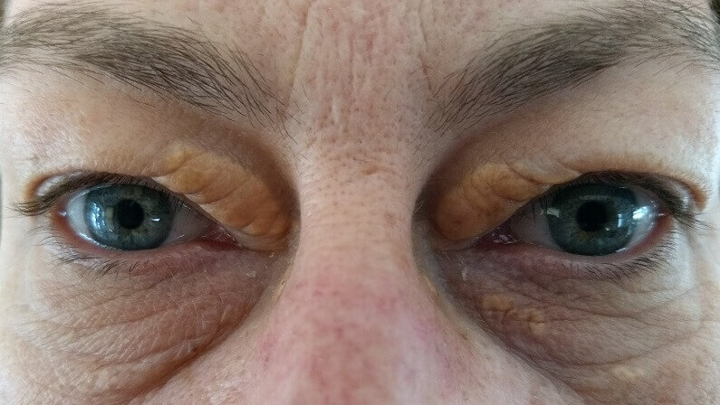
Soft, yellow to yellow-orange patches with fairly clear edges, usually on the upper lid near the inner corner and often on both sides. They can be flat or slightly raised and range from tiny spots to plaques over a centimeter long.
The inner corner (medial canthus) is the classic site, and eyelid xanthomas are more common on the upper lid than the lower.[2] They’re painless, don’t itch and don’t affect your vision. A doctor can usually diagnose them just by looking.[2]
Over months and years they may grow, thicken and merge. Our guides to what xanthelasma looks like, xanthelasma on the eyelid and xanthelasma of the eyelid show the range of appearances.
How Is An Eyelid Xanthoma Different From Xanthomas Elsewhere?
Eyelid xanthomas are common and often harmless, and about half occur with normal blood fats. Xanthomas on tendons, as sudden crops of bumps or as widespread flat patches are less common and more strongly linked to significant blood fat or blood protein disorders.
Tendon xanthomas, firm lumps on the Achilles tendons, hands, elbows and knees, are a classic sign of familial hypercholesterolemia, an inherited condition that keeps LDL cholesterol high from birth.[4] Widespread flat plane xanthomas, especially with normal cholesterol, have been linked to monoclonal gammopathy, a blood protein disorder.[5] Eyelid xanthomas can occur alongside these, including as part of familial hypercholesterolemia,[4] but most often they appear on their own.
That’s why an eyelid-only xanthoma is usually a routine matter, while xanthomas elsewhere always deserve a doctor’s workup. Our guides to tendon xanthomas, eruptive xanthoma and plane xanthoma cover the other types.

Eyelid Xanthoma And Other Xanthomas Compared
| Type | Where | Often Linked To | What To Do |
|---|---|---|---|
| Eyelid xanthoma (xanthelasma) | Eyelids, inner corners | Blood fat disorder in about half; often cosmetic | Routine check-up; removal if wanted |
| Tendon xanthoma | Achilles, hands, elbows, knees | Familial hypercholesterolemia | See a doctor; lipid and family assessment |
| Eruptive xanthoma | Buttocks, arms, legs | Very high triglycerides | Prompt medical attention |
| Diffuse plane xanthoma | Neck, trunk, skin folds, sometimes eyelids | Blood protein disorders, often with normal lipids | Doctor’s workup with blood protein tests |
What Can An Eyelid Xanthoma Signal?
About half of people with an eyelid xanthoma have a blood fat disorder, such as raised LDL or low HDL, and some have diabetes, an underactive thyroid or a liver condition. It’s also been linked with a higher risk of heart disease.
About 50% of people with xanthelasma have a primary or secondary lipid disorder.[1] The American Academy of Ophthalmology notes that 25% to 70% have normal blood fats when first seen, and lists associations with diabetes, an underactive thyroid, heart disease, obesity and smoking.[2] In younger people, doctors consider inherited high cholesterol.[1]
The usual check is a fasting lipid profile, liver panel, thyroid tests and blood sugar.[1] Our guide to the causes of xanthelasma explains each one.

Why Do Xanthomas Form On The Eyelids?

Blinking squeezes the tiny blood vessels in the thin eyelid skin, and these vessels become leakier with age. Fat escapes into the skin, immune cells swallow it, and they swell into foam cells that cluster into a plaque.
Researchers think this local process explains why the eyelids are the most common site for xanthomas, and why they can form even when blood fats are normal.[1] Higher blood fats simply mean more fat available to leak. Our guide on how xanthelasma forms goes step by step.

Can You Have Eyelid Xanthomas And Xanthomas Elsewhere?
Yes, and when you do, it’s more significant. Eyelid xanthomas together with tendon lumps are a classic pattern in familial hypercholesterolemia, and eyelid plaques alongside widespread flat patches can point to a blood protein disorder.
Familial hypercholesterolemia, which affects about 1 in 250 to 1 in 500 people and passes to each child with a 50% chance, can cause both xanthelasma and tendon xanthomas on the Achilles tendons, hands, elbows, knees and feet. Without medical care, about half of men with it had a coronary event by 50, so finding it matters.[4] Diffuse plane xanthomas can involve the eyelids as well as the neck and trunk, and have been linked to monoclonal gammopathy.[5]
It’s worth a quick self-check: run your fingers along the back of your heels and over your knuckles for firm lumps, and look for yellow patches on your palms, neck or body. If you find anything, tell your doctor, as it changes which tests they’ll want to do.
- Eyelids plus tendon lumps. Ask about inherited high cholesterol and family testing.
- Eyelids plus widespread flat patches. Ask about blood protein tests.
- Eyelids only. A routine check-up and blood test is usually enough.

When Is An Eyelid Xanthoma Not Simple Xanthelasma?
Rarely, yellow eyelid lesions are part of a different condition. Necrobiotic xanthogranuloma causes firm yellow-orange plaques that can ulcerate, and it’s strongly linked to blood protein disorders. Some rare histiocytic conditions also cause xanthelasma-like plaques.
Necrobiotic xanthogranuloma (NXG) produces firm, yellow-to-orange papules, nodules or plaques, most often around the eyes, which can ulcerate and scar. Between 77% and 84% of people with NXG eventually develop a blood disorder, most commonly a paraproteinemia, and doctors recommend a biopsy and a blood protein test.[3] It usually begins around the sixth decade of life.[3]
Other rarities include Erdheim-Chester disease, in which about 30% of people have xanthelasma-like plaques, and syringomas, small skin-colored bumps that can be confused with xanthelasma.[2] Widespread flat plane xanthomas can also involve the eyelids.[5] The clues are firmness, ulceration, rapid spread or lesions elsewhere on the body. Our guide on whether xanthelasma can be cancerous covers these rare lookalikes.

How Is An Eyelid Xanthoma Diagnosed?
Usually just by looking. A doctor then arranges blood tests for cholesterol, liver, thyroid and blood sugar. A biopsy is only needed if the lesion looks unusual.
- Examination. The look and position are usually enough to confirm xanthelasma.[2]
- Blood tests. Fasting lipid profile, liver panel, thyroid tests and fasting glucose.[1]
- Family history. Relatives with high cholesterol or early heart disease suggest an inherited cause.[4]
- Biopsy if unusual. Firm, ulcerated or atypical lesions are biopsied.[1][3]
- Blood protein tests if needed. Recommended when necrobiotic xanthogranuloma is suspected.[3]

How Do You Remove A Xanthoma From The Eyelid?
With laser, surgery, a chemical peel or radiofrequency at a clinic, or a removal cream made for xanthelasma at home. Lowering cholesterol protects your health but rarely clears existing plaques.
A review of 49 studies found CO2 laser gave more than 75% improvement in over 90% of patients, and Er:YAG laser more than 80%, with skin color change the most common complication; surgery is used for larger, deeper plaques.[6] Surgery can change the eyelid’s position, and freezing and chemical methods can scar or discolor the skin.[1] Our guides to the removal of xanthelasma and xanthelasma cream compare the options.
Once a doctor has confirmed your eyelid xanthoma is ordinary xanthelasma and checked your blood, this is the at-home cream we make for removing it.

Will An Eyelid Xanthoma Come Back After Removal?
It can. Recurrence after removal is generally under 50%, higher for deeper plaques and after repeated procedures, and has been reported at 13% to 16% after laser.
Recurrence is generally under 50%, and deeper plaques come back more often.[1] Laser recurrence has been reported at 13% to 16%.[2] Managing any cholesterol, thyroid or blood sugar problem gives you the best chance of fewer new plaques. Our guide on whether xanthelasma comes back covers this.

When Should You See A Doctor About An Eyelid Xanthoma?
Once for any new eyelid xanthoma, to confirm it and check your blood. Promptly if it’s firm, ulcerated, bleeding or spreading fast, or if you have xanthomas elsewhere on your body.
A Routine Visit Is Fine If
- The plaques are soft, yellow and on the eyelids only
- They’re slow to change
- You’d like a cholesterol check
- You’re thinking about removal
See A Doctor Promptly If
- A plaque is firm, ulcerated or scarring
- Plaques are spreading quickly
- You have lumps on tendons, elbows or knees
- Flat yellow patches appear on your body
Our guide to the xanthoma overview explains how all the types fit together.
What’s The Bottom Line On Eyelid Xanthomas?
A xanthoma on the eyelid is xanthelasma, the most common and usually simplest type of xanthoma. About half of people have a blood fat disorder behind it, so have one check-up; firm or ulcerated lesions need a closer look.
Once a doctor has confirmed it, removal is a cosmetic choice. Xanthel® is a cosmetic cream made for xanthelasma removal at home, sold by Xanthelasma.com, for the eyelid type only. You’ll find it at the foot of this page, or read more about Xanthel® xanthelasma removal cream and browse all our xanthelasma guides.
Common Questions About Eyelid Xanthomas
Is An Eyelid Xanthoma The Same As Xanthelasma?
Yes. Xanthoma is the family name for fat-filled deposits, and xanthelasma is the name for one on the eyelids.
Is An Eyelid Xanthoma Dangerous?
Usually not. It’s harmless in itself, but about half of people have a blood fat disorder, and rare firm or ulcerated lesions can be a different condition.
Does An Eyelid Xanthoma Mean I Have High Cholesterol?
Not necessarily. About half of people with one have a blood fat disorder and about half have normal results, so a blood test is the only way to know.
Are Eyelid Xanthomas Different From Other Xanthomas?
Yes. Eyelid xanthomas are common and often occur with normal cholesterol, while tendon, eruptive and widespread flat xanthomas point more strongly to significant underlying disorders.
Can An Eyelid Xanthoma Be Something Else?
Rarely. Necrobiotic xanthogranuloma, linked to blood protein disorders, can cause firm yellow-orange eyelid plaques that ulcerate, so unusual lesions need a biopsy.
Will An Eyelid Xanthoma Go Away On Its Own?
Rarely. The fat is locked inside cells in the skin, so it usually stays or slowly grows unless it’s removed.
Can An Eyelid Xanthoma Be Removed At Home?
Yes, once a doctor has confirmed it’s ordinary xanthelasma. An at-home cream such as Xanthel®, applied only to the plaque, is one way to remove it.
Should I See A Doctor About An Eyelid Xanthoma?
Yes, once, to confirm it and check your cholesterol, thyroid, liver and blood sugar, and promptly if it’s firm, ulcerated or spreading.
Sources
- Blair K. Xanthelasma Palpebrarum. StatPearls, National Library of Medicine (NCBI Bookshelf), updated 2026.
- Patadia A, Rana HS, Stewart K, Yen MT, Lee H. Xanthelasma. EyeWiki, American Academy of Ophthalmology, reviewed 2026.
- Kamboj A, Santilli C, Yamanuha J, Harrison A, Maltry A, Hou J, Mokhtarzadeh A, Vercellotti G. Necrobiotic Xanthogranuloma. EyeWiki, American Academy of Ophthalmology, updated 2026.
- Ison HE, Clarke SL, Knowles JW. Familial Hypercholesterolemia. GeneReviews, National Library of Medicine (NCBI Bookshelf), revised 2025.
- Szalat R, et al. Pathogenesis and treatment of xanthomatosis associated with monoclonal gammopathy. Blood, 2011.
- Malekzadeh H, Ormseth B, Janis JE. A Practical Review of the Management of Xanthelasma Palpebrarum. Plastic and Reconstructive Surgery Global Open, 2023;11(5):e4982.