What The Word Means, Where It Comes From, Every Type Defined, And Where Eyelid Xanthelasma Fits In
A xanthoma is a yellowish deposit of fat-filled cells in the skin, tendons or another tissue. The word comes from the Greek for “yellow”, and it’s an umbrella term covering several types, from eruptive bumps and tendon swellings to xanthelasma, the soft yellow plaques on the eyelids. Most are harmless in themselves but can signal a blood fat disorder or another condition.
Xanthoma definition: a localized yellowish deposit of lipid inside tissue, usually the skin or tendons, made of immune cells filled with fat (foam cells). Types include eruptive, tuberous, tendinous, plane (planar) xanthomas and xanthelasma on the eyelids. They’re usually benign but often point to a lipid disorder or other condition.
Key Takeaways
- A xanthoma is a local deposit of lipid, usually in the skin or tendons.
- The yellow color comes from fat-filled immune cells called foam cells.
- The plural is xanthomas or xanthomata.
- Main types: eruptive, tuberous, tendinous, plane, and xanthelasma.
- Most are benign but can signal a blood fat disorder or other illness.
- Xanthelasma is the eyelid type and can be removed at home or in a clinic.
On This Page 14 sections
What Is The Definition Of A Xanthoma?
A xanthoma is a localized deposit of lipid within a tissue, most often the skin, the tissue just under it, or a tendon. It looks yellow or orange because it’s made of immune cells packed with fat.
Medically, xanthomas are local lipid deposits within an organ system. They’re usually benign in themselves, but they often signal a systemic condition, most commonly one affecting blood fats.[1] That’s the key to the definition: a xanthoma is a sign as much as a lesion, and the type and place tell a doctor a lot about what’s behind it.
“Xanthoma” is an umbrella term. Eruptive xanthomas, tendon xanthomas, palmar xanthomas and eyelid xanthelasma are all xanthomas, but they look different, sit in different places and point to different things. Our main xanthomas guide covers them in depth, and what is xanthoma gives a quick introduction.

Where Does The Word Xanthoma Come From, And How Do You Say It?
It comes from the Greek “xanthos”, meaning yellow, and “-oma”, a suffix used for a growth or swelling. It’s pronounced zan-THOH-muh, and the plural is xanthomas or xanthomata.
- Xanthos. Greek for yellow, the color of the deposits.
- -oma. A suffix used for a growth or swelling, as in many medical terms. It doesn’t mean cancer here.
- Pronunciation. zan-THOH-muh, with the “x” sounding like a “z”.
- Plural. Xanthomas in everyday English, or xanthomata, the older Greek-style plural. See our guide to xanthomata.
The same root gives xanthelasma, from “xanthos” and “elasma”, a plate, which is why it’s often described as yellow plaques. Our guide on how to pronounce xanthelasma covers that word too.

What Do The Related Terms Mean?
Xanthomatous means “relating to or like a xanthoma”, xanthomatosis means having many xanthomas, and xanthelasma is the eyelid type. Foam cells are the fat-filled immune cells that make up every xanthoma.
- Foam cell. An immune cell (macrophage) that has taken up so much lipid it looks foamy under a microscope.[1]
- Xanthomatous. Describes something with the features of a xanthoma. See xanthomatous.
- Xanthomatosis. A condition with many xanthomas, often widespread. See xanthomatosis.
- Xanthelasma (palpebrarum). Soft yellow plaques on the eyelids, the best-known type.[2]
- Plane (planar) xanthoma. A flat xanthoma, for example in the palm creases or skin folds.[1] See plane xanthoma.
- Xanthoma disseminatum. A rare condition with widespread xanthoma-like lesions. See xanthoma disseminatum.
You’ll see these words used together in clinic letters and test reports. A doctor might write that a patient has “bilateral xanthelasma and Achilles tendon xanthomas, suggesting familial hypercholesterolemia”, or describe a skin biopsy as showing “xanthomatous change with foam cells”. In each case the meaning is the same: yellow, fat-filled cells have gathered in the tissue, and the next question is why.

How Does A Xanthoma Form?
When blood fats are high, fat-carrying particles leak through the walls of small blood vessels into the skin or tendons. Immune cells swallow the fat and turn into foam cells, which gather to form a yellow deposit.
In people with raised blood fats, lipoproteins seep through the lining of blood vessels into the dermis, the tissue under the skin and tendons, and tissue macrophages engulf the lipid to become foam cells.[1] Xanthomas can also form with normal blood fats: injury or inflammation can make vessels leakier, which is thought to explain lesions at sites of frequent mechanical trauma.[1]
That second route matters for xanthelasma. Blinking compresses the eyelid’s small vessels, which become leakier with age, so lipid can escape even when blood levels are normal.[2]

What Are The Types Of Xanthoma?
The main types are eruptive, tuberous and tuberoeruptive, tendinous, plane (planar), and xanthelasma on the eyelids. Rarer forms are linked with blood protein disorders and inherited conditions such as cerebrotendinous xanthomatosis.
- Eruptive. Small inflamed yellow-red bumps that appear in crops within days and fade over weeks.[1] See eruptive xanthoma.
- Tuberous and tuberoeruptive. Firmer red to yellow papules that can merge, favoring the elbows and palms.[1] See tuberous xanthoma.
- Tendinous. Slowly growing swellings in the Achilles tendons, elbows and the extensor tendons of the hands, strongly linked with familial hypercholesterolemia.[1] See tendon xanthomas.
- Plane (planar). Flat deposits in the palm creases, flexures and skin folds; palmar crease xanthomas can be pathognomonic for familial dysbetalipoproteinemia.[1] See palmar xanthoma.
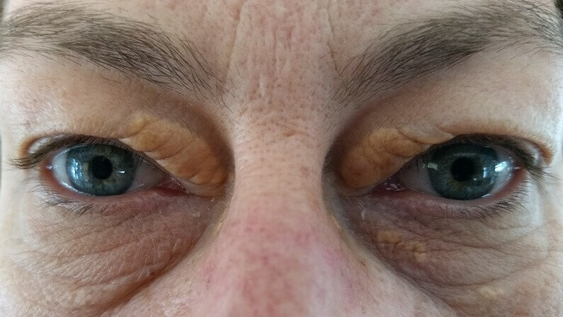
- Xanthelasma. Soft yellow plaques on the eyelids, usually near the inner corners.[2]
- Paraprotein-related. Flat, grouped lesions that can occur with normal blood fats.[1]
- Cerebrotendinous xanthomatosis. A rare inherited condition affecting the brain and tendons.[1]

The Types Of Xanthoma Compared
| Type | Where | What It Looks Like | Often Linked With |
|---|---|---|---|
| Xanthelasma | Eyelids, near the inner corners | Soft, flat or slightly raised yellow plaques | A lipid disorder in about half of people; normal blood fats in the rest |
| Eruptive | Often in crops on the body | Crops of small yellow-red bumps | Very high triglycerides, uncontrolled diabetes |
| Tuberous | Elbows, knees, palms | Firm nodules that can merge | Inherited lipid disorders |
| Tendinous | Achilles, elbows, hand tendons | Slowly growing tendon swellings | Familial hypercholesterolemia |
| Plane (palmar) | Palm creases, skin folds | Flat yellow-orange lines or patches | Dysbetalipoproteinemia, cholestatic liver disease |
| Paraprotein-related | Body, often widespread | Flat, grouped patches | Monoclonal gammopathy, normal blood fats |
Where Does Xanthelasma Fit Into The Xanthoma Definition?
Xanthelasma is the eyelid type of xanthoma: soft yellow plaques near the inner corners of the eyes. It’s the best-known type, it’s linked with a lipid disorder in only about half of people, and it’s usually a cosmetic concern.
Xanthelasma palpebrarum affects about 1.1% of women and 0.3% of men, usually between ages 20 and 70, and about half of people with it have a primary or secondary lipid disorder.[2] In a Swiss study of 5,504 benign eyelid skin tumors, about 6% were xanthelasmas.[1]
Unlike tendon or palmar xanthomas, which are strong markers of particular inherited disorders, xanthelasma often appears with normal blood fats. It’s still worth a check-up, because it’s linked with a lipid disorder in about half of people.[2] Our main xanthelasma guide and what xanthelasma looks like cover the eyelid type in full.

What Do Xanthomas Indicate?

Often a blood fat disorder, but the type matters. Tendon xanthomas point to familial hypercholesterolemia, palmar ones to dysbetalipoproteinemia, eruptive ones to very high triglycerides, and some flat xanthomas with normal blood fats to a blood protein disorder.
- Familial hypercholesterolemia. About 75% of older adults with it have tendon xanthomas.[1] It affects about 1 in 250 to 1 in 500 people, and each child of an affected parent has a 50% chance of inheriting it.[3]
- Dysbetalipoproteinemia. An inherited disorder in which cholesterol and triglycerides both rise; it causes palmar and tuberous xanthomas.[4]
- Very high triglycerides. Eruptive xanthomas appear with triglycerides above 1,000 mg/dL, often with uncontrolled diabetes, and carry a risk of pancreatitis.[5]
- Other conditions. Diabetes, an underactive thyroid and kidney disease (nephrotic syndrome) can raise blood fats and lead to xanthomas.[1]
- Blood protein disorders. Diffuse flat xanthomas with normal blood fats have been linked with monoclonal gammopathy.[6]
The prognosis depends mainly on the underlying condition rather than the xanthoma itself.[1] Our guide to xanthoma explains each link in more detail.

How Is A Xanthoma Diagnosed?
Usually by examination, followed by blood tests for the cause: fasting triglycerides and cholesterol, plus blood sugar and HbA1c where needed. Tendon xanthomas may need ultrasound or MRI, and a biopsy is used if the diagnosis is uncertain.
- Examination. A clinical exam is usually enough to recognize a xanthoma.[1]
- Fasting blood tests. Triglycerides, total, LDL and HDL cholesterol, with glucose and HbA1c as indicated.[1]
- Pattern-specific checks. Palmar crease or diffuse plane xanthomas prompt a search for their associated conditions.[1]
- Imaging for tendons. Ultrasound can assess tendon xanthomas, and MRI helps rule out other causes and plan surgery.[1]
- Eyelid workup. For xanthelasma: a fasting lipid profile, liver panel, thyroid tests and fasting glucose.[2]
- Biopsy. Used to rule out other diagnoses when needed.[1]

How Are Xanthomas Managed?
Mainly by managing the condition behind them, usually with cholesterol-lowering medicine and lifestyle changes. Small xanthomas often shrink or clear once that’s under control; larger or troublesome ones can be removed surgically.
Controlling the underlying condition is the main goal. Statins lower blood fats and may shrink the lesions, and PCSK9 inhibitor injections are effective in familial hypercholesterolemia.[1] Small xanthomas often shrink or resolve with good control, and some have cleared after liver transplant or plasma exchange.[1] In dysbetalipoproteinemia, palmar crease xanthomas fade with time once management begins.[4]
Surgery is used for tendon xanthomas that rub on footwear, ideally combined with lipid control to lower the chance of them returning.[1] Xanthelasma is the exception to the “fades with control” rule: managing the cause rarely clears eyelid plaques, which is why they’re usually removed directly (see below).

How Is Eyelid Xanthelasma Removed?
With clinic procedures such as surgery, laser or chemical peels, or at home with a removal cream made for xanthelasma. Plaques can return, with recurrence generally under 50%.
Clinic options carry their own risks: surgical excision can make the lower lid turn outward or pull back, and freezing and chemical methods can scar and discolor the skin. Recurrence is generally under 50%, rising to around 60% after repeat procedures.[2] Many people with small to medium eyelid plaques prefer to remove them at home, and our xanthelasma cream guide explains how that works.
If you have the eyelid type, your doctor has checked the cause, and you want to deal with the plaques themselves at home, this is the cream we make for xanthelasma. It’s for eyelid xanthelasma only, not for xanthomas elsewhere on the body.

When Should You See A Doctor About A Xanthoma?
Whenever you find one. Every type is worth a blood test, and some need prompt attention: sudden crops of bumps, swellings on tendons, or yellow lines in the palm creases.
- Eyelid plaques. Book a routine check for blood fats, thyroid, liver and blood sugar.
- Tendon swellings or palm-crease lines. Ask about inherited lipid disorders and family testing.
- Sudden crops of yellow-red bumps. See a doctor soon; very high triglycerides carry a pancreatitis risk.[5]
- Severe upper stomach pain. Get urgent help, as it can be a sign of pancreatitis.
- Widespread flat patches with normal blood tests. Ask whether a blood protein disorder has been considered.[6]
Our guide to what xanthomas are helps you describe what you’ve found.

What’s The Bottom Line On The Xanthoma Definition?
A xanthoma is a yellow deposit of fat-filled foam cells in the skin or tendons, named from the Greek for yellow. The type tells you most: tendon, palmar and eruptive xanthomas point to specific lipid disorders, while eyelid xanthelasma often occurs with normal blood fats.
Whatever the type, a blood test is the first step. If yours is eyelid xanthelasma and you want it gone, Xanthel® is a cosmetic cream made for xanthelasma removal at home, sold by Xanthelasma.com, for the eyelid type only. You’ll find it at the foot of this page, or read more about Xanthel® xanthelasma removal cream, and browse all our xanthelasma and xanthoma guides.
Common Questions About The Xanthoma Definition
What Is The Definition Of A Xanthoma?
A xanthoma is a localized yellowish deposit of lipid in a tissue, usually the skin or tendons, made of fat-filled immune cells called foam cells.
What Does The Word Xanthoma Mean?

It comes from the Greek “xanthos”, meaning yellow, and “-oma”, a suffix for a growth or swelling. It doesn’t mean cancer.
What Is The Plural Of Xanthoma?
Xanthomas in everyday English, or xanthomata, the older Greek-style plural.
Is Xanthelasma A Xanthoma?
Yes. Xanthelasma is the eyelid type of xanthoma, and about half of people with it have normal blood fats.
What Are The Main Types Of Xanthoma?
Eruptive, tuberous, tendinous, plane (including palmar) and xanthelasma, plus rarer types linked with blood protein disorders and inherited conditions.
Is A Xanthoma Cancer?
No. Xanthomas are benign deposits of fat-filled cells, though they can signal a condition elsewhere in the body.
Do Xanthomas Go Away?
Small ones often shrink or clear once the underlying condition is controlled. Eyelid xanthelasma rarely clears that way and is usually removed directly.
Should I See A Doctor About A Xanthoma?
Yes. Every type is worth a blood test, and sudden crops of bumps, tendon swellings or palm-crease lines need prompt attention.
Sources
- Bell A, Shreenath AP. Xanthoma. StatPearls, National Library of Medicine (NCBI Bookshelf), updated 2023.
- Blair K. Xanthelasma Palpebrarum. StatPearls, National Library of Medicine (NCBI Bookshelf), updated 2026.
- Ison HE, Clarke SL, Knowles JW. Familial Hypercholesterolemia. GeneReviews, National Library of Medicine (NCBI Bookshelf), revised 2025.
- Javvaji A, Can AS, Sharma S. Dysbetalipoproteinemia. StatPearls, National Library of Medicine (NCBI Bookshelf), updated 2024.
- Mishra C, Tripathy K. Lipemia Retinalis. StatPearls, National Library of Medicine (NCBI Bookshelf), updated 2026.
- Szalat R, et al. Pathogenesis and treatment of xanthomatosis associated with monoclonal gammopathy. Blood, 2011.