How To Tell Xanthelasma From Other Eyelid Lumps, The Rare Eyelid Xanthomas Worth Knowing, And What To Do Once You Know Which You Have
Eyelid xanthomas are yellow fatty deposits in the eyelid skin, and almost all of them are xanthelasma palpebrarum: soft, yellow plaques near the inner corners, often on both eyes. A few rarer conditions and several ordinary eyelid bumps can look similar, so the first step is knowing which one you have.
Eyelid xanthomas are usually xanthelasma: soft, yellow, flat or slightly raised plaques near the inner eyelid corners, often on both sides. They’re painless and don’t affect sight, but about half of people have a blood fat disorder. Firm, orange, spreading or sore lesions may be something else and need a doctor. Xanthelasma plaques can be removed.
Key Takeaways
- Most eyelid xanthomas are xanthelasma palpebrarum, the commonest xanthoma.
- Soft, yellow, flat plaques near the inner corners are the classic sign.
- Milia, styes, chalazia and syringomas are common lookalikes.
- Firm, orange or spreading plaques can signal rarer conditions needing a biopsy.
- About half of people with xanthelasma have a lipid disorder worth testing for.
- Xanthelasma can be removed; rarer eyelid xanthomas need specialist care.
On This Page 16 sections
What Are Eyelid Xanthomas?
Eyelid xanthomas are deposits of fat-filled immune cells in the eyelid skin. The common form is xanthelasma palpebrarum, the most frequent type of xanthoma anywhere on the body; much rarer types also affect the area around the eyes.
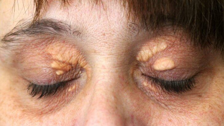
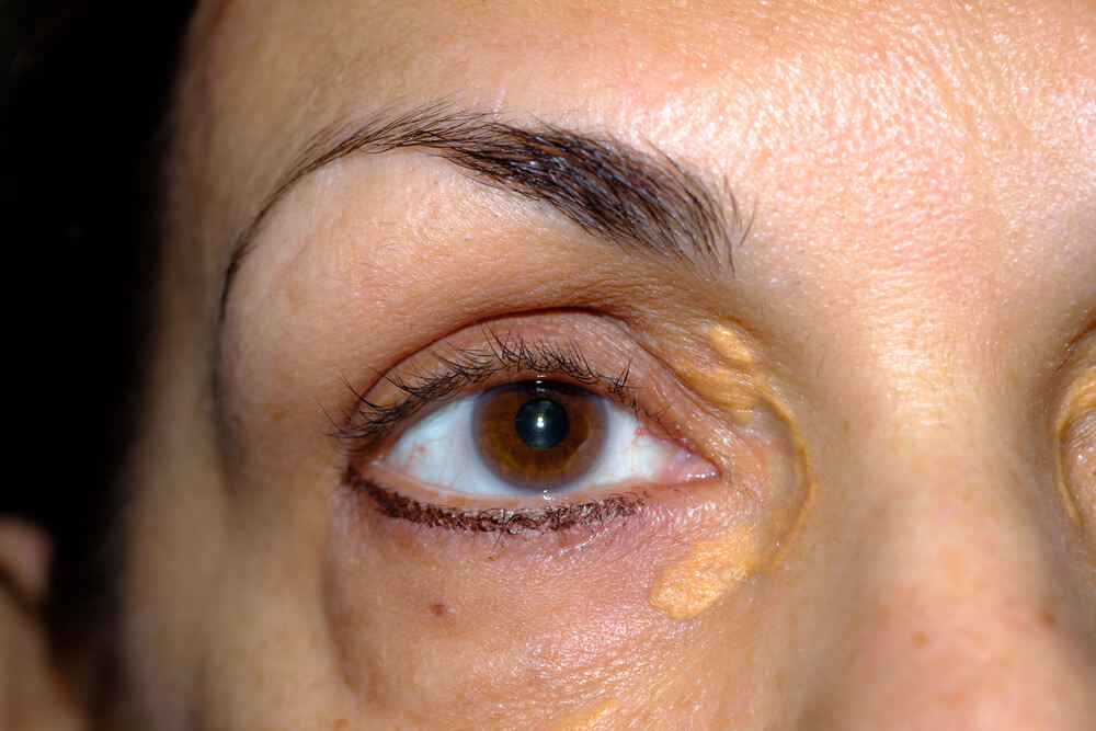
A xanthoma is a collection of lipid-laden immune cells, called foam cells, in the skin or tendons. Xanthelasma palpebrarum is the most common xanthoma, and it appears as soft, yellow plaques on the eyelids, usually near the inner corners.[1] So when a doctor or website talks about “eyelid xanthomas,” they almost always mean xanthelasma, the cholesterol deposits on the eyelids that most people notice in the mirror.
Our main xanthelasma guide covers the condition in depth, and our xanthomas guide explains the wider family. For a shorter introduction to the eyelid form, see xanthoma eyelid.

What Do Eyelid Xanthomas Look Like?
Soft, yellow to yellow-orange plaques, flat or slightly raised, with clear edges, usually near the inner corners of the eyelids and often matching on both sides. They’re more common on the upper lid.
- Color. Pale cream to yellow, sometimes orange-tinged; most other eyelid bumps are skin-colored, white or red.
- Texture. Soft or velvety rather than hard.
- Shape. A flat patch or plaque, not a tense round dome.
- Position. Near the inner corner, more often on the upper lid, frequently on both sides.[2]
- Size. A small dot to larger patches that can join together.
Our guides on what xanthelasma looks like, whether it can be flat and whether it can be white show the range of appearances.

Why Do Xanthomas Form On The Eyelids?
Blinking repeatedly squeezes the tiny eyelid blood vessels, which become leakier with age. Fats seep into the skin, immune cells swallow them and become foam cells, and the foam cells collect into plaques.
The foam cells gather in the deeper part of the skin, the reticular dermis, around the vessels, with some inflammation.[1] About half of people with xanthelasma have a primary or secondary lipid disorder; secondary causes include diabetes, an underactive thyroid, primary biliary cholangitis, nephrotic syndrome and retinoid or estrogen medicines.[1] In others, blood fats are normal and local eyelid factors seem to matter more. Our guide to the causes of xanthelasma explains each one.

Who Gets Eyelid Xanthomas?
Mostly adults in midlife, and women more than men. Xanthelasma affects about 1.1% of women and 0.3% of men, usually between 20 and 70 and most often between 35 and 55.
Those figures come from StatPearls, which also advises considering an inherited lipid disorder when xanthelasma appears in young people.[1] Inherited high cholesterol, such as familial hypercholesterolemia, can cause both eyelid xanthomas and lumps on the tendons. Our guide on whether xanthelasma is genetic covers who’s most at risk, and how common it is.
What Other Eyelid Lumps Look Like Eyelid Xanthomas?
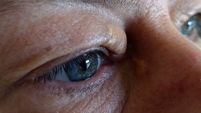
Milia, styes, chalazia, skin tags, syringomas and sebaceous hyperplasia are the common lookalikes. Rarely, a skin cancer such as basal cell carcinoma can appear on the eyelid, which is why unusual spots need checking.
- Milia. Tiny, hard, white or pearly dots, much smaller than xanthelasma.
- Stye. A red, tender lump at the lash line that appears quickly.
- Chalazion. A firmer, rounder lump from a blocked eyelid gland, usually single and not yellow.
- Skin tags. Small, soft, flesh-colored flaps.
- Syringoma. Small skin-colored or yellowish bumps that often cluster under the eyes.[3]
- Sebaceous hyperplasia. Small yellowish bumps, often with a central dip.[3]
- Nodular basal cell carcinoma. A pearly bump, sometimes with tiny vessels; needs a doctor.[3]
Our guide on what can be mistaken for xanthelasma goes through each lookalike with pictures.

Eyelid Lump Checker
| Lump | Color And Feel | Where And How | What To Do |
|---|---|---|---|
| Xanthelasma (eyelid xanthoma) | Yellow, soft, flat or slightly raised | Inner corners, often both eyes; grows slowly | Doctor’s check and blood tests; removal if wanted |
| Milia | White, tiny, hard | Anywhere on the lids or cheeks | Usually harmless; ask if unsure |
| Stye | Red, tender | At the lash line; appears quickly | Warm compresses; see a doctor if it worsens |
| Chalazion | Skin-colored, firm, round | Within the lid; usually single | See a doctor if it persists |
| Syringoma | Skin-colored or yellowish, small, firm | Clusters, often under the eyes | Dermatologist if unsure |
| Basal cell carcinoma | Pearly, may bleed or crust | Single, slowly enlarging | See a doctor promptly |
| Necrobiotic xanthogranuloma | Yellow to orange or violet, firm | Spreading around the eyes | See a doctor; biopsy and blood tests |
What Are The Rare Types Of Eyelid Xanthoma?
Necrobiotic xanthogranuloma, adult-onset asthma with periocular xanthogranuloma (AAPOX), Erdheim-Chester disease and widespread plane xanthoma can all cause yellow plaques around the eyes. They’re rare, but unlike xanthelasma they’re linked with conditions that need specialist care.
EyeWiki lists necrobiotic xanthogranuloma, Erdheim-Chester disease and AAPOX among the conditions to rule out when eyelid plaques look like xanthelasma.[2] Necrobiotic xanthogranuloma causes yellowish or violaceous plaques around the eyes and is linked with blood protein disorders.[3] Diffuse plane xanthomas in people with normal blood fats have been linked with monoclonal gammopathy, an abnormal blood protein.[5] These are uncommon, and most people with eyelid xanthomas have ordinary xanthelasma, but they’re the reason firm, spreading or unusual plaques deserve a proper look. Our guide to plane xanthoma covers the flat type in more detail.

Which Eyelid Lumps Need A Doctor Promptly?
See a doctor promptly for any eyelid lump that’s painful, bleeding, crusting or ulcerated, pearly with tiny vessels, firm and orange or violet, spreading around the eye, changing fast, or causing loss of lashes.
- Bleeding, crusting or a sore that won’t heal. Could be a skin cancer.
- Firm, orange or violet plaques spreading around the eyes. Could be a rarer xanthoma.
- Pain, redness or swelling. Suggests infection or inflammation.
- A single lump growing quickly. Needs assessment.
- Loss of lashes over a lump. A warning sign on the lid margin.

Our guide on whether xanthelasma can be cancerous explains the differences.

How Does A Doctor Confirm Eyelid Xanthomas?
Usually just by looking. Typical xanthelasma is a clinical diagnosis. A biopsy is used when the appearance is unusual or a rarer condition is suspected, and blood tests check for the cause.
Under the microscope, xanthelasma shows foamy macrophages in the deeper skin with the surface layer spared.[2] The usual blood tests are a fasting lipid profile, liver panel, thyroid tests and fasting glucose, with HbA1c in diabetes.[1] If a rare type is suspected, a doctor may add blood protein tests. Our guide on which doctor to see explains who does what.
What Do Eyelid Xanthomas Mean For Your Health?
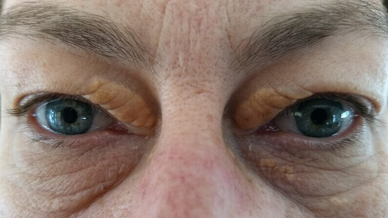
They’re a reason to check your blood fats and heart health. About half of people with xanthelasma have a lipid disorder, and xanthelasma is linked with a higher risk of heart attack and heart disease, even with normal cholesterol.
A study that followed almost 13,000 people for decades found that those with xanthelasma had a higher risk of heart attack and heart disease, independent of their cholesterol.[6] The plaques themselves sit in the eyelid skin, don’t hurt and don’t affect your sight; the health message is about what may be happening elsewhere. Our guide on what xanthelasma indicates explains the tests and results.

How Are Eyelid Xanthomas Different From Xanthomas On The Body?
Eyelid xanthelasma is usually a soft, flat plaque that may or may not come with a lipid disorder. Xanthomas on the tendons, palms, elbows or buttocks are more often a direct sign of a specific lipid disorder and are dealt with mainly by getting that disorder under control with a doctor.
For example, tendon xanthomas on the Achilles, elbows, hands or knees are a sign of familial hypercholesterolemia, which also causes xanthelasma. Our guides to tendon xanthomas, skin xanthomas and xanthoma care by type explain the body forms.

Do Eyelid Xanthomas Change Over Time?
They tend to stay the same or slowly grow, and new plaques can appear or join together. They rarely fade by themselves, even when cholesterol is brought down.
Xanthelasma doesn’t typically resolve on its own, and once plaques have formed, lifestyle changes and lipid-lowering medicine may have only a limited effect on how they look.[1] Plaques that suddenly appear, change fast or become firm aren’t typical. Our guides on how fast xanthelasma grows and whether it can go away on its own explain what to expect.

Can Eyelid Xanthomas Be Removed?
Yes, once a doctor has confirmed typical xanthelasma. Clinic options are laser, surgery and acid peels; small to medium plaques can also be removed at home with a cream made for xanthelasma. Rare eyelid xanthomas need specialist care instead.
A 2023 review of 49 studies found CO2 laser gave more than 75% improvement in over 90% of patients and erbium laser more than 80%, with changes in skin color the most common complication; surgery was favored for larger, deeper plaques.[4] Freezing is generally avoided near the eye because of swelling.[2] Many people with smaller plaques prefer xanthelasma removal at home. Our guides to the removal of xanthelasma and Xanthel cream compare the options.
If your doctor has confirmed your eyelid xanthomas are xanthelasma and you want to deal with them at home, this is the cream we make for it.

Will Eyelid Xanthomas Come Back After Removal?
They can. After removal, xanthelasma comes back in under half of people overall, around 60% after repeat procedures, and more often with deeper plaques. Managing the cause lowers the odds.
Recurrence varies by method and depth.[1] Keep up any cholesterol, thyroid or blood sugar care your doctor recommends, check your eyelids every few months, and deal with new spots while they’re small. Our guides on whether xanthelasma comes back and how you stop xanthelasma explain how.

What’s The Bottom Line On Eyelid Xanthomas?

Eyelid xanthomas are almost always xanthelasma: soft, yellow plaques near the inner corners. Get them confirmed and have your blood fats checked, watch for firm, spreading or unusual plaques that may be rarer types, and remove typical xanthelasma in a clinic or at home if it bothers you.
Xanthel® is a cosmetic cream made for xanthelasma removal at home, sold by Xanthelasma.com, for eyelid xanthelasma only. You’ll find it in the box above, or read more about Xanthel® xanthelasma removal cream, and browse all our xanthelasma guides.
Common Questions About Eyelid Xanthomas
What Are Eyelid Xanthomas?
Deposits of fat-filled immune cells in the eyelid skin. Almost all are xanthelasma palpebrarum, soft yellow plaques near the inner corners.
Are Eyelid Xanthomas The Same As Xanthelasma?
Usually, yes. Xanthelasma is the common eyelid xanthoma, but rare conditions such as necrobiotic xanthogranuloma can also cause yellow plaques around the eyes.
How Do I Know If My Eyelid Lump Is A Xanthoma?
Xanthelasma is soft, yellow, flat or slightly raised and near the inner corner, often on both eyes. Firm, red, painful, pearly or bleeding lumps are something else.
What Else Could A Yellow Bump On My Eyelid Be?
Syringoma, sebaceous hyperplasia or milia are common lookalikes, and rarely a skin cancer or a rarer xanthogranuloma, so unusual spots should be checked.
Are Eyelid Xanthomas Dangerous?
The plaques themselves don’t hurt or affect sight, but about half of people with xanthelasma have a lipid disorder and it’s linked with higher heart risk, so get checked.
Do Eyelid Xanthomas Mean High Cholesterol?
Not always. About half of people with xanthelasma have a lipid disorder; the rest have normal blood fats. A blood test is the only way to know.
Can Eyelid Xanthomas Be Removed?
Typical xanthelasma can be removed with laser, surgery or peels in a clinic, or at home with a cream made for xanthelasma. Rare types need specialist care.
Will Eyelid Xanthomas Come Back?
They can. Xanthelasma returns in under half of people after removal, more often with deeper plaques, so keep managing the cause.
Sources
- Blair K. Xanthelasma Palpebrarum. StatPearls, National Library of Medicine (NCBI Bookshelf), updated 2026.
- Patadia A, Rana HS, Stewart K, Yen MT, Lee H. Xanthelasma. EyeWiki, American Academy of Ophthalmology, reviewed September 2026.
- Van Alfen B, Hatch J, Conley M, Seeliyur Duraiswamy S. A Case Report of Xanthelasma and the Associated Differential Diagnosis. Cureus, 2025;17(4):e81971.
- Malekzadeh H, Ormseth B, Janis JE. A Practical Review of the Management of Xanthelasma Palpebrarum. Plastic and Reconstructive Surgery Global Open, 2023;11(5):e4982.
- Szalat R, et al. Pathogenesis and treatment of xanthomatosis associated with monoclonal gammopathy. Blood, 2011.
- Christoffersen M, Frikke-Schmidt R, Schnohr P, Jensen GB, Nordestgaard BG, Tybjaerg-Hansen A. Xanthelasmata, arcus corneae, and ischaemic vascular disease and death in general population: prospective cohort study. BMJ, 2011.