Why The Plaques Are Benign, Which Rare Conditions Can Look Similar, And The Warning Signs Worth Checking
Can xanthelasma be cancerous? No. Xanthelasma is a benign buildup of fat-filled immune cells in the eyelid skin, and it isn’t cancer or a precancerous growth. A few rare conditions, including one eyelid cancer, can look similar, which is why a doctor should confirm the diagnosis once. What xanthelasma more often points to is a cholesterol problem, not cancer.
No, xanthelasma isn’t cancerous. It’s a harmless collection of fat-filled immune cells in the eyelid skin. Rarely, an eyelid cancer called sebaceous carcinoma, or conditions linked to blood disorders, can cause yellowish eyelid lumps, so hard, ulcerated, bleeding or fast-growing patches need a doctor’s check.
Key Takeaways
- Xanthelasma is benign: it isn’t cancer and isn’t precancerous.
- It’s made of fat-filled immune cells, not abnormal growing cells.
- Sebaceous carcinoma, a rare eyelid cancer, can also look yellow.
- Necrobiotic xanthogranuloma looks similar and is linked to blood disorders.
- Hard, ulcerated, bleeding or fast-growing patches need a prompt check.
- Xanthelasma’s main health link is to cholesterol and the heart.
On This Page 14 sections
Can Xanthelasma Be Cancerous?
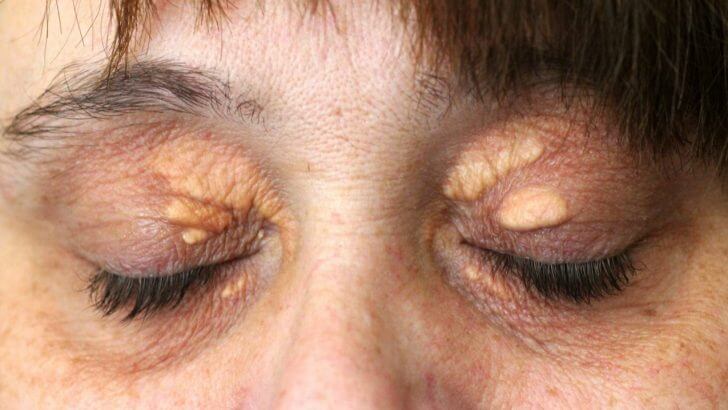
No. Xanthelasma is a benign condition: a collection of fat-filled immune cells in the eyelid skin. It isn’t a tumor, it doesn’t spread to other parts of the body, and it isn’t a sign of cancer.
Xanthelasma palpebrarum is the most common type of xanthoma, a family of fatty deposits in the skin and tendons, and it’s described in the medical literature as benign.[1] It affects around 1.1% of women and 0.3% of men, usually in midlife.[1] Plaques can grow slowly and new ones can appear, which sometimes worries people, but that’s the fat deposit building up, not cancer spreading.
The main reason to see a doctor about xanthelasma isn’t cancer at all: it’s that about half of people with it have a blood fat disorder.[1] Our guides on whether xanthelasma is dangerous and whether xanthelasma spreads explain more, and our main guide to xanthelasma covers the whole condition.
Why Isn’t Xanthelasma Cancer?
Because it’s made of ordinary immune cells that have filled with fat, not abnormal cells growing out of control. The plaque forms when these cells collect fat that has leaked from tiny eyelid blood vessels.
Cancer happens when cells grow and divide abnormally, invade nearby tissue and can spread. Xanthelasma is different. Blinking squeezes the small blood vessels in the eyelid, and as they become leakier with age, fat escapes into the skin. Immune cells called macrophages swallow it, swell into foam cells, and gather around the vessels with some inflammation.[1]
So the yellow plaque is a collection of normal cells doing a clean-up job and becoming overloaded with fat. That’s why it looks yellow, why it’s soft, and why it doesn’t behave like a tumor. Our guides to what xanthelasma is and how xanthelasma forms go into the details.
Can Xanthelasma Turn Into Cancer Over Time?
No. Xanthelasma isn’t a precancerous condition. It can grow, thicken and spread to nearby skin over the years, but it stays a benign fat deposit.
Plaques often start small and flat, then slowly enlarge or merge, and older plaques tend to be firmer and deeper. That’s a change in size and depth, not a change into something malignant. Many people live with xanthelasma for decades with no change other than slow growth.
What a doctor will want to confirm is that what you have really is xanthelasma in the first place. If a “xanthelasma” plaque starts behaving unusually, such as becoming hard, ulcerating, bleeding or growing quickly, that’s a reason to have it looked at, because it may be something else.
Is Xanthelasma Ever Linked To Cancer?
Ordinary xanthelasma isn’t. But two rarer conditions that can look like it are linked to blood disorders: necrobiotic xanthogranuloma, and widespread flat (plane) xanthomas with normal cholesterol. Both have been associated with conditions such as monoclonal gammopathy and multiple myeloma.
Necrobiotic xanthogranuloma (NXG) causes firm, yellow-to-orange papules, nodules or plaques, most often around the eyes, which can ulcerate and scar. Between 77% and 84% of people with NXG eventually develop a blood disorder, most often a paraproteinemia (an abnormal blood protein), and progression to multiple myeloma has been reported up to six years after the skin changes appear.[3] It usually starts in the sixth decade of life, and doctors recommend a blood protein test and a biopsy.[3]
Diffuse plane xanthomas, flat yellow patches spreading over the neck, trunk or skin folds, often with normal cholesterol, have been linked with monoclonal gammopathy.[5] Some rare histiocytic disorders can also cause xanthelasma-like plaques; for example, about 30% of people with Erdheim-Chester disease have them.[2] Our guides to plane xanthoma and xanthomas explain these conditions.

Which Eyelid Cancers Can Look Like Xanthelasma?
The main one is sebaceous carcinoma, a rare cancer of the eyelid’s oil glands. It often looks yellow, usually affects the upper lid, and can be mistaken for a stye or blepharitis. Basal cell carcinoma, the most common eyelid cancer, usually looks different.
Sebaceous carcinoma is a rare but aggressive skin cancer that most often arises in the eyelid’s oil glands. It’s typically yellow because the tumor cells contain fat, often has irregular borders, and is more common on the upper lid. The reported median age at diagnosis is 57 to 72.[4] It can mimic a chalazion (a blocked oil gland) or chronic blepharitis, and warning signs include a recurring or unusual chalazion on the upper lid, yellow thickening of the lid, and eyelid inflammation that won’t settle.[4]

It’s rare: in one large review of eyelid tumors, sebaceous carcinoma made up 3% of malignant cases, while basal cell carcinoma made up 86%.[4] Early diagnosis matters, because in a large analysis it had spread in about 12% of cases.[4] The key differences from xanthelasma are firmness, thickening of the lid itself and involvement of the lid edge. Our guides to what can look like xanthelasma and what can be mistaken for xanthelasma cover the full list.

Xanthelasma And Its Serious Lookalikes Compared
| Condition | How It Looks And Feels | Linked To | What To Do |
|---|---|---|---|
| Xanthelasma | Soft, yellow, flat or slightly raised; inner corners; both sides | Blood fat disorder in about half; not cancer | Routine check-up and blood test; removal if wanted |
| Necrobiotic xanthogranuloma | Firm, yellow-orange, can ulcerate and scar; around the eyes | Blood protein disorders, sometimes myeloma | Prompt specialist review, biopsy, blood protein test |
| Sebaceous carcinoma | Yellow thickening or lump, often upper lid, may involve the lid edge | Eyelid cancer | Prompt eye specialist review and biopsy |
| Diffuse plane xanthoma | Flat yellow patches spreading beyond the eyelids | Monoclonal gammopathy, often with normal lipids | Doctor’s workup with blood protein tests |
What Warning Signs Should You Look Out For?
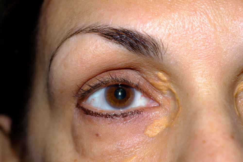
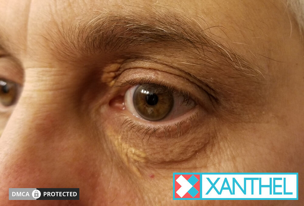
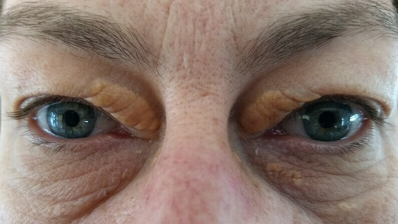
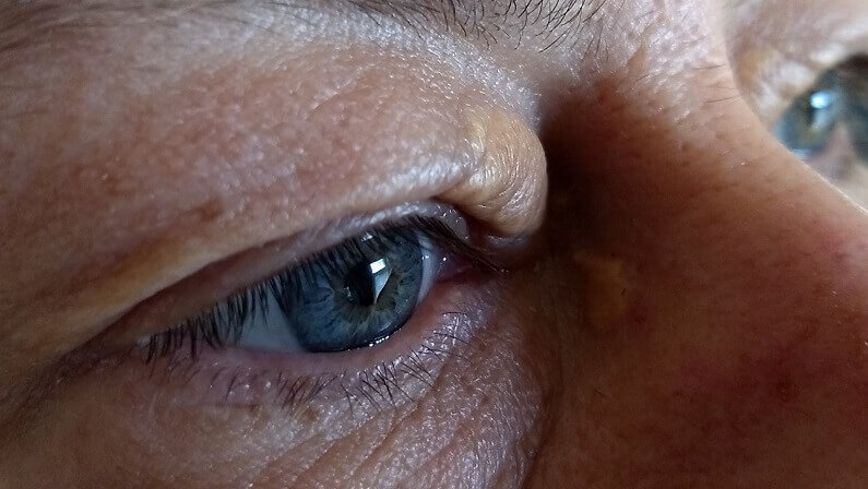
Typical xanthelasma is soft, yellow, painless, at the inner corners, on both sides and slow to change. Anything hard, ulcerated, bleeding, fast-growing, involving the lid edge or spreading beyond the eyelids deserves a prompt check.
Typical Of Xanthelasma
- Soft and smooth
- Inner corners of the eyelids
- Similar on both sides
- Slow, steady change over years
Worth A Prompt Check
- Hard, ulcerated, crusting or bleeding
- Growing quickly or changing shape
- Thickening of the lid edge or a lump that keeps coming back
- Yellow patches spreading beyond the eyelids
Most of the time, a check-up simply confirms xanthelasma. But if anything feels unusual, it’s always better to ask. Our guide to what xanthelasma looks like shows the typical appearances, and can xanthelasma be white covers color changes.
How Do Doctors Tell Xanthelasma From Something More Serious?
Usually just by looking: typical xanthelasma is easy to recognize. If anything is unusual, a small biopsy confirms the diagnosis, and blood tests check cholesterol and, where needed, blood proteins.
- Examination. A doctor can usually diagnose xanthelasma by its look and position.[1][2]
- Blood tests. A fasting lipid profile, liver panel, thyroid tests and blood sugar for xanthelasma.[1]
- Biopsy if unsure. A small sample shows foam cells in xanthelasma, and different features in lookalikes.[1][3]
- Blood protein test if needed. Serum protein electrophoresis is recommended when necrobiotic xanthogranuloma is suspected.[3]
- Specialist review. A suspicious eyelid lump goes to an eye specialist for biopsy.[4]

What Does Xanthelasma Mean For Your Health, If Not Cancer?
Its main health link is to cholesterol and the heart. About half of people with xanthelasma have a blood fat disorder, and xanthelasma has been linked with a higher risk of heart attack and heart disease.
About 50% of people with xanthelasma have a primary or secondary lipid disorder, such as raised LDL or low HDL, or another condition such as diabetes, an underactive thyroid or a liver problem.[1] A Danish study that followed almost 13,000 people for decades found that people with xanthelasma had a higher risk of heart attack and heart disease, regardless of their cholesterol levels.[6]
So the useful check-up for xanthelasma is a blood test, not a cancer scan. Our guides to what xanthelasma indicates and whether xanthelasma indicates raised cholesterol explain what your results might show.

Is It Safe To Remove Xanthelasma?
Yes, once a doctor has confirmed it’s xanthelasma. Removal is cosmetic, and there’s no cancer risk in removing a benign plaque. Clinic methods carry some risk of scarring and color change, and at-home removal is an alternative.
Because xanthelasma is benign, removing it is a choice about appearance and comfort, not a medical necessity. Clinic options include laser, surgery, chemical peels and radiofrequency; surgery can change the position of the eyelid, and freezing or chemical methods can scar or discolor the skin.[1] Recurrence after removal is generally under 50%.[1] Our guides to the removal of xanthelasma, what doctor removes xanthelasma and xanthelasma cream compare the options.
Once your doctor has confirmed your plaques are xanthelasma and checked your blood, if you’d like to remove them at home, this is the cream we make for xanthelasma.

When Should You See A Doctor?
Once for any new yellow eyelid patches, to confirm the diagnosis and check your cholesterol. Promptly if a patch is hard, ulcerated, bleeding, growing fast, involves the lid edge or spreads beyond the eyelids.
Your family doctor is the right first stop and can refer you to a dermatologist or eye specialist if anything looks unusual. If an eyelid lump has been called a chalazion but keeps coming back, or the eyelid stays inflamed despite care, ask about a biopsy.[4] For typical xanthelasma, one routine visit with a blood test is usually all you need before deciding whether to remove it.
What Can You Do If You’re Still Worried About Cancer?
Keep a simple record of your plaques, see your doctor if anything changes, and don’t be afraid to ask for a biopsy if a patch looks or behaves differently from typical xanthelasma.
- Take clear photos. Photograph your eyelids in daylight every month or two, from the same angle.
- Note the details. Write down the size, color and feel of each patch, and whether both sides match.
- Watch for change. Slow, steady growth is typical; sudden change, hardness, bleeding or crusting isn’t.
- Mention your history. Tell your doctor about any recurring styes, eyelid inflammation or blood disorders.
- Ask the direct question. It’s perfectly reasonable to ask, “Could this be anything other than xanthelasma, and would a biopsy help?”
Worry is common with anything on the face, and a clear answer from a doctor is usually reassuring. Most of the time, it simply confirms xanthelasma.
What’s The Bottom Line On Whether Xanthelasma Can Be Cancerous?
Xanthelasma isn’t cancer and doesn’t turn into cancer. It’s a benign buildup of fat-filled cells. A few rare lookalikes are more serious, so have a doctor confirm the diagnosis once, especially if a patch is hard, ulcerated, bleeding or fast-growing.
The more useful check for xanthelasma is a cholesterol test, because about half of people with it have a blood fat disorder. Once you know it’s xanthelasma, removal is simply a cosmetic choice. Xanthel® is a cosmetic cream made for xanthelasma removal at home, sold by Xanthelasma.com. You’ll find it at the foot of this page, or read more about Xanthel® xanthelasma removal cream and browse all our xanthelasma guides.
Common Questions About Xanthelasma And Cancer
Can Xanthelasma Be Cancerous?
No. Xanthelasma is a benign collection of fat-filled immune cells in the eyelid skin. It isn’t cancer and isn’t a precancerous condition.
Can Xanthelasma Turn Into Cancer?
No. It can grow, thicken and spread to nearby skin over time, but it stays a benign fat deposit.
Is Xanthelasma A Sign Of Cancer?
Ordinary xanthelasma isn’t. Rare lookalikes, such as necrobiotic xanthogranuloma and widespread plane xanthomas, are linked to blood disorders, which is why unusual patches deserve a check.
What Eyelid Cancer Looks Like Xanthelasma?
Sebaceous carcinoma, a rare cancer of the eyelid’s oil glands, can look yellow. It’s usually a firmer thickening or lump, often on the upper lid, and can be mistaken for a stye.
How Can I Tell If My Xanthelasma Is Something Else?
Typical xanthelasma is soft, yellow, painless and at the inner corners on both sides. Hard, ulcerated, bleeding or fast-growing patches should be checked promptly.
Do I Need A Biopsy For Xanthelasma?
Usually not. A doctor can normally diagnose xanthelasma by looking, and a biopsy is only needed if the appearance is unusual.
What Does Xanthelasma Mean For My Health?
Its main link is to cholesterol and the heart. About half of people with it have a blood fat disorder, so a blood test is the most useful check.
Is It Safe To Remove Xanthelasma?
Yes, once a doctor has confirmed it’s xanthelasma. Removal is cosmetic, done at a clinic or at home, and carries no cancer risk.
Sources
- Blair K. Xanthelasma Palpebrarum. StatPearls, National Library of Medicine (NCBI Bookshelf), updated 2026.
- Patadia A, Rana HS, Stewart K, Yen MT, Lee H. Xanthelasma. EyeWiki, American Academy of Ophthalmology, reviewed 2026.
- Kamboj A, Santilli C, Yamanuha J, Harrison A, Maltry A, Hou J, Mokhtarzadeh A, Vercellotti G. Necrobiotic Xanthogranuloma. EyeWiki, American Academy of Ophthalmology, updated 2026.
- Silverstein E, Marcet MM. Sebaceous Carcinoma. EyeWiki, American Academy of Ophthalmology, reviewed 2026.
- Szalat R, et al. Pathogenesis and treatment of xanthomatosis associated with monoclonal gammopathy. Blood, 2011.
- Christoffersen M, Frikke-Schmidt R, Schnohr P, Jensen GB, Nordestgaard BG, Tybjaerg-Hansen A. Xanthelasmata, arcus corneae, and ischaemic vascular disease and death in general population: prospective cohort study. BMJ, 2011.