A Plain-English Introduction To Xanthomas: What These Yellow Fatty Deposits Are, How They Form, The Main Types, And What They Can Tell You About Your Health
Xanthomas are yellowish deposits of fat in the skin or around tendons, made of fat-filled cells called foam cells. They’re noncancerous and usually painless, but they often signal raised blood fats, an inherited cholesterol condition, diabetes or a thyroid problem. The eyelid type, xanthelasma, is the best known; other types appear on the elbows, knees, hands, feet, tendons and buttocks.
A xanthoma is a yellow to orange fatty deposit in the skin or around a tendon. They form when fat leaks into tissue and immune cells swallow it. Types include eruptive, tendon, tuberous, palmar, plane and eyelid xanthomas (xanthelasma). They’re harmless in themselves but can point to high cholesterol, very high triglycerides or diabetes, so see a doctor.
Key Takeaways
- Xanthomas are noncancerous, usually painless fatty deposits in the skin or tendons.
- They often signal raised blood fats or another condition worth checking.
- Eruptive xanthomas come from very high triglycerides, which can lead to pancreatitis.
- Tendon xanthomas often point to familial hypercholesterolemia.
- Eyelid xanthelasma is the most familiar type; about half of people with it have a lipid disorder.
- A doctor can usually recognize them on sight and confirm the cause with blood tests.
On This Page 17 sections
What Is Xanthomas?
Xanthomas (the plural of xanthoma) are yellowish deposits of fat that build up in the skin or around tendons. They’re noncancerous and painless, but they can be a sign of another condition, most often a problem with blood fats.
The word comes from the Greek for “yellow”, which describes the color most of them share. MedlinePlus describes xanthomas as common, especially in older adults and people with high blood lipids, varying in size and able to appear anywhere on the body, most often on the elbows, joints, tendons, knees, hands, feet or buttocks.[1]
“Xanthoma” isn’t one condition but a family of related deposits. Some come up in crops over days; others grow slowly over years. Some sit on the skin’s surface; others lie deep around tendons. What links them is what they’re made of, and the fact that they’re often a visible clue to what’s happening in your blood.
For the full guide to every type, see our pillar page on xanthomas. Our pages on what a xanthoma is and the xanthoma definition cover the single lesion and the medical terms.

How Do Xanthomas Form?
Fat-carrying particles leak out of small blood vessels into the surrounding tissue. Immune cells called macrophages swallow the fat, become overloaded and turn into foam cells, which cluster into the yellow deposit you can see.
StatPearls describes this process for eyelid xanthomas: blinking constantly squeezes the tiny vessels in the eyelid, vessels get leakier with age, and macrophages overloaded with fat become foam cells around the vessels in the deeper skin, with some inflammation.[5] The same basic step, fat escaping into tissue and being taken up by macrophages, underlies xanthomas elsewhere.
The more fat-carrying particles there are in the blood, the more can escape. That’s why very high triglycerides can bring on crops of eruptive xanthomas, and why lifelong high LDL cholesterol in inherited conditions can build up deposits around tendons. Our page on xanthomata explains how the plural forms are described.

What Do Xanthomas Look Like?
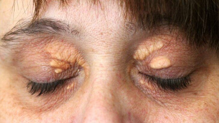
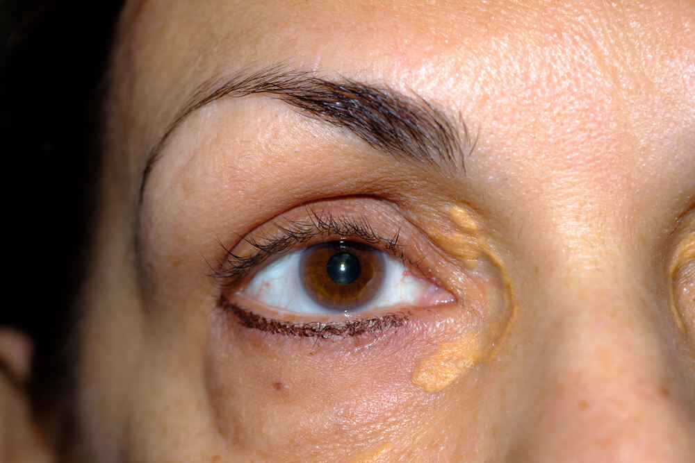
Usually yellow to orange bumps or patches with clear edges, alone or in clusters. Some are flat, some raised, some firm nodules, and those around tendons may not change the skin color at all.
MedlinePlus notes that some xanthomas are yellow to orange bumps with defined borders, either single or in clusters, while those next to tendons don’t change the color of the skin.[1] Eruptive xanthomas look different again: pea-sized, waxy, firm bumps from yellow to red, sometimes with a small red halo, which may itch or feel tender.[2]
On The Skin
- Yellow, orange or reddish color
- Flat patches or raised bumps
- Clear edges, single or clustered
- Soft on the eyelids, firmer elsewhere
Around Tendons
- Firm lumps under the skin
- Often skin-colored, not yellow
- Common on the Achilles and knuckles
- Move with the tendon
Our guides to skin xanthomas and xanthoma on the skin show how they appear in more detail.

What Are The Main Types Of Xanthomas?
Eruptive, tendon, tuberous, palmar, plane and eyelid xanthomas (xanthelasma). They’re named by their shape and where they appear, and each tends to point to a different cause.
Doctors sort xanthomas by how they look and where they sit, because that’s often the quickest clue to what’s behind them. A crop of red-yellow bumps on the buttocks suggests very high triglycerides; a firm lump on the Achilles tendon suggests inherited high cholesterol; yellow lines in the palm creases suggest a specific inherited lipid disorder. The sections below take each type in turn, with our guide to xanthomatosis covering what it means when many appear at once.

What Are Eruptive Xanthomas?
Crops of pea-sized, waxy, yellow to red bumps on the buttocks, shoulders, arms, thighs and legs, caused by very high triglycerides, often with poorly controlled diabetes.
MedlinePlus describes eruptive xanthomatosis as a rare condition caused by an excessively high triglyceride level. It can occur with poorly controlled diabetes, and medicines such as isotretinoin, estrogens and cyclosporine can trigger it.[2] The bumps usually go away by themselves after a few weeks once blood sugar and fat levels are under control, but high triglycerides left unmanaged can lead to pancreatitis.[2] Our page on eruptive xanthoma explains more.

What Are Tendon Xanthomas?

Firm lumps on tendons, most often the Achilles, knuckles, elbows and knees, that usually point to familial hypercholesterolemia or, rarely, another inherited condition.
Familial hypercholesterolemia affects about 1 in 250 to 1 in 500 people, and each child of an affected parent has a 50% chance of inheriting it. It often causes xanthelasma and tendon xanthomas on the Achilles tendons, elbows, hands, knees and feet, and, left unmanaged, about 50% of men had a coronary event by 50 and about 30% of women by 60.[3]
A rarer cause is cerebrotendinous xanthomatosis, an inherited condition in which tendon xanthomas appear in the teens to twenties, often after childhood diarrhea and cataracts. Recognizing it early matters, because early care with a bile acid medicine appears to prevent symptoms.[6] Our guide to tendon xanthomas covers both.

What Are Tuberous And Palmar Xanthomas?
Tuberous xanthomas are firm, yellowish nodules over the elbows and knees; palmar xanthomas are yellow streaks in the creases of the palms. Both are characteristic of dysbetalipoproteinemia, an inherited lipid disorder.
StatPearls describes palmar and tuberous xanthomas as characteristic of dysbetalipoproteinemia, in which cholesterol- and triglyceride-rich particles build up in the blood, and notes that palmar xanthomas fade once the condition is managed.[4] Tuberous xanthomas can also appear with familial hypercholesterolemia. Our pages on tuberous xanthoma and palmar xanthoma describe each.

What Are Plane Xanthomas?
Flat yellow patches that can spread over the neck, trunk, skin folds or around the eyes. Some come with a lipid disorder; others appear with normal blood fats and deserve a thorough check-up.
Because xanthomas can be linked with a range of conditions, including certain cancers as well as lipid disorders,[1] widespread flat patches with normal cholesterol are a reason to see a doctor rather than simply living with them. Our guide to plane xanthoma explains what doctors look for.

What Are Eyelid Xanthomas (Xanthelasma)?
Soft, flat or slightly raised yellow plaques near the inner corners of the eyelids. Xanthelasma is the most familiar xanthoma, and about half of people with it have a lipid disorder.
Xanthelasma palpebrarum affects about 1.1% of women and 0.3% of men, most often between 35 and 55. About 50% of people with it have a primary or secondary lipid disorder, so StatPearls recommends a fasting lipid profile, liver panel, thyroid tests and fasting glucose.[5] These cholesterol deposits on the eyelids rarely fade on their own, and they’re the one type of xanthoma that can be removed at home as well as at a clinic.
Our guides to xanthelasma, xanthoma on the eyelid and what causes cholesterol deposits around the eyes go into the eyelid type in depth.

If yours is the eyelid type and you’re managing the cause with your doctor, this is the cream we make for removing eyelid plaques at home.
Types Of Xanthomas At A Glance
| Type | Looks Like | Where | Often Linked With |
|---|---|---|---|
| Eruptive | Crops of pea-sized, waxy yellow to red bumps | Buttocks, shoulders, arms, thighs, legs | Very high triglycerides, poorly controlled diabetes |
| Tendon | Firm, often skin-colored lumps | Achilles, knuckles, elbows, knees | Familial hypercholesterolemia |
| Tuberous | Firm yellowish nodules | Elbows, knees | Dysbetalipoproteinemia, high cholesterol |
| Palmar | Yellow streaks in skin creases | Palms and fingers | Dysbetalipoproteinemia |
| Plane | Flat yellow patches | Neck, trunk, skin folds, around the eyes | Lipid disorders, sometimes other conditions |
| Eyelid (xanthelasma) | Soft, flat yellow plaques | Inner corners of the eyelids | A lipid disorder in about half |
What Can Xanthomas Signal?
Raised cholesterol or triglycerides, an inherited lipid disorder, diabetes, an underactive thyroid, liver or pancreatic conditions and, rarely, certain cancers.
MedlinePlus lists conditions that raise blood lipids and can bring on xanthomas: certain cancers, diabetes, high blood cholesterol, familial hypercholesterolemia, primary biliary cholangitis (formerly primary biliary cirrhosis), pancreatitis and an underactive thyroid.[1] For eyelid plaques, StatPearls adds nephrotic syndrome, retinoid or estrogen medicines and, in young people, inherited lipid disorders.[5]
- Heart and arteries. High LDL, especially when inherited, raises the risk of early heart disease.[3]
- Pancreas. Very high triglycerides can lead to pancreatitis.[2]
- Blood sugar. Poorly controlled diabetes can drive eruptive xanthomas.[2]
- Family. Inherited conditions mean relatives may benefit from testing too.[3]
Our page on the causes of xanthelasma goes through the eyelid causes one by one.

Who Gets Xanthomas?

They’re common, especially in older adults and people with high blood fats, but inherited conditions can cause them in children and young adults too.
MedlinePlus describes xanthomas as common, particularly in older adults and people with high blood lipids.[1] Eyelid xanthelasma is more common in women and most often appears between 35 and 55.[5] Inherited conditions shift things younger: in cerebrotendinous xanthomatosis, tendon xanthomas appear in the teens to twenties,[6] and in young people with eyelid plaques, an inherited lipid disorder is more likely.[5] People with poorly controlled diabetes are at risk of eruptive xanthomas.[2]

Are Xanthomas Dangerous?
The deposits themselves aren’t cancerous or harmful, but what they signal can be: high cholesterol that raises heart risk, or very high triglycerides that can lead to pancreatitis.
MedlinePlus describes xanthomas as noncancerous and painless, while noting they may indicate another medical condition.[1] That’s the key point: think of a xanthoma as a warning light rather than a problem in itself. Some, such as eruptive xanthomas, can itch or feel tender,[2] and tendon lumps can rub on shoes, but the real reason to act is the health check they prompt.

How Are Xanthomas Diagnosed?
Usually by a doctor looking at the skin, with a small biopsy if needed, plus blood tests for cholesterol, triglycerides, liver function and diabetes to find the cause.
- A look at the skin. Doctors usually recognize xanthomas by examining them.[1]
- Blood tests. Lipid levels, liver function and diabetes checks.[1]
- Thyroid and pancreatic tests. Where the picture suggests them.[2]
- A skin biopsy. A tiny sample if the diagnosis isn’t clear.[1]
- Family and genetic testing. If an inherited condition is suspected.[3]
Doctors also rule out spots that only resemble xanthomas. Our page on what can be mistaken for xanthelasma covers the eyelid lookalikes.

What Should You Do If You Think You Have Xanthomas?
See your doctor for a skin check and blood tests, manage whatever is found, and then decide whether any remaining lesion needs removing. Get prompt care for sudden crops of bumps.
Managing the underlying condition may help reduce new xanthomas, and a doctor can remove a bothersome one surgically or with a laser, though it may return.[1] Eruptive xanthomas usually clear within weeks once triglycerides and blood sugar are under control,[2] and palmar xanthomas fade once the lipid disorder is managed.[4]
Xanthomas on the body, hands, elbows, knees or tendons should be left to a doctor. Only eyelid xanthelasma can also be removed at home. Our guide to how each type of xanthoma is managed walks through the options.
What’s The Bottom Line On What Xanthomas Are?
Xanthomas are noncancerous, yellowish fatty deposits in the skin or around tendons, formed when fat leaks into tissue and is swallowed by immune cells. They come in several types and often signal raised blood fats, an inherited lipid disorder or diabetes, so a doctor’s check is the right first step.
Xanthel® is a cosmetic cream made for xanthelasma removal at home, sold by Xanthelasma.com. It’s for eyelid xanthelasma only, not xanthomas on the body. You’ll find it in the box above and at the foot of this page, or read more about Xanthel® xanthelasma removal cream. You can also browse all our xanthelasma and xanthoma guides.
Common Questions About Xanthomas
What Is A Xanthoma?
A xanthoma is a yellowish, noncancerous deposit of fat in the skin or around a tendon, made of fat-filled immune cells called foam cells.
What Is The Difference Between Xanthoma And Xanthelasma?
Xanthoma is the general term for these fatty deposits; xanthelasma is the type that appears on the eyelids.
What Causes Xanthomas?
Usually raised blood fats, from high cholesterol, very high triglycerides, inherited lipid disorders, diabetes, an underactive thyroid or liver conditions.
What Are The Types Of Xanthomas?
The main types are eruptive, tendon, tuberous, palmar, plane and eyelid xanthomas, also called xanthelasma.
Are Xanthomas Dangerous?
The deposits aren’t cancerous, but they can signal high cholesterol that raises heart risk or very high triglycerides that can lead to pancreatitis.
Do Xanthomas Mean High Cholesterol?

Often, but not always. About half of people with eyelid xanthelasma have a lipid disorder, and blood tests are the only way to know.
Can Xanthomas Be Removed?
Yes. A doctor can remove a bothersome xanthoma surgically or with a laser, and eyelid xanthelasma can also be removed with a cream at home.
Should I See A Doctor About Xanthomas?
Yes. Any new xanthoma may signal a condition that needs care, so a skin check and blood tests are worthwhile.
Sources
- Xanthoma. MedlinePlus Medical Encyclopedia, National Library of Medicine, reviewed April 2025.
- Eruptive xanthomatosis. MedlinePlus Medical Encyclopedia, National Library of Medicine, reviewed October 2024.
- Ison HE, Clarke SL, Knowles JW. Familial Hypercholesterolemia. GeneReviews, National Library of Medicine (NCBI Bookshelf), revised 2025.
- Javvaji A, Can AS, Sharma S. Dysbetalipoproteinemia. StatPearls, National Library of Medicine (NCBI Bookshelf), updated 2024.
- Blair K. Xanthelasma Palpebrarum. StatPearls, National Library of Medicine (NCBI Bookshelf), updated 2026.
- Federico A, Gallus GN. Cerebrotendinous Xanthomatosis. GeneReviews, National Library of Medicine (NCBI Bookshelf), revised 2024.