Reading The Signs On Your Skin: A Body Map Of Xanthomas, What Each Type Looks Like, Which Blood Problem It Points To, And What To Do About It
A skin xanthoma is a yellowish deposit of fat-filled cells in the skin, and where it appears says a lot about what’s behind it. Soft plaques on the eyelids are xanthelasma, the most common type; firm lumps on tendons point to inherited high cholesterol; sudden crops of small bumps point to very high triglycerides; and widespread flat patches with normal cholesterol need checking for a blood disorder. Every new xanthoma deserves a blood test.
Skin xanthomas are yellow deposits of fat-filled immune cells in the skin. Types include xanthelasma on the eyelids, tendon, tuberous, palmar, eruptive and plane xanthomas. Each points to different blood fat problems, from inherited high cholesterol to very high triglycerides, so every new one deserves a doctor’s check and blood tests.
Key Takeaways
- A skin xanthoma is a deposit of fat-filled immune cells, called foam cells.
- Xanthelasma on the eyelids is the most common type.
- Tendon xanthomas are a classic sign of familial hypercholesterolemia.
- Eruptive xanthomas can appear when triglycerides top 1,000 mg/dL.
- Body xanthomas often shrink when blood fats are controlled; eyelid ones rarely do.
- Every new xanthoma deserves a doctor’s check and blood tests.
On This Page 16 sections
What Is A Skin Xanthoma?
A yellow, orange or skin-colored deposit in the skin made of immune cells packed with fat. It can be a flat patch, a soft plaque, a firm lump or a crop of small bumps, depending on the type and what’s driving it.
The name comes from the Greek xanthos, meaning yellow. Xanthomas are a sign rather than a disease in themselves: they show that fats from the blood have built up in the skin or tendons, and the pattern often points to a particular lipid problem. The best-known example is xanthelasma on the eyelids, which StatPearls, the National Library of Medicine’s clinical reference, places at about 1.1% of women and 0.3% of men.[1]
Our pages on xanthoma, what xanthoma is and skin xanthomas give the background. This page is a map: where each type appears and what it’s telling you.

How Do Xanthomas Form In The Skin?
Fat-carrying particles leak from small blood vessels into the skin, immune cells called macrophages swallow them, and the cells become foam cells that gather into a visible deposit.
StatPearls describes this process for xanthelasma: the foam cells collect around small vessels in the deeper skin layer, with some inflammation around them.[1] The same basic mechanism applies elsewhere; what differs is which fats are raised, how high, and where in the body they settle. Pressure and movement seem to matter too, which is why many types favor eyelids, elbows, knees, tendons and skin creases.
Because the fat is held inside living cells, xanthomas can’t be squeezed out. Our page on xanthomatosis covers what happens when many xanthomas appear at once.

Where On The Skin Do Xanthomas Appear?
Eyelids, tendons, elbows and knees, palm creases, buttocks and the backs of arms and legs, and sometimes broad areas of the neck and trunk. The site is one of the best clues to the type.
- Eyelids, inner corners. Xanthelasma: soft, flat or slightly raised yellow plaques.[1]
- Achilles tendons, knuckles, elbows, knees. Tendon xanthomas: firm lumps that move with the tendon.[2]
- Elbows, knees, buttocks. Tuberous xanthomas: firm yellow nodules over pressure points.[3]
- Palm and finger creases. Palmar xanthomas: yellow lines in the creases.[3]
- Buttocks, arms, legs. Eruptive xanthomas: sudden crops of small yellow-red bumps.[4]
- Eyelids, neck, trunk, folds. Plane xanthomas: flat yellow patches, sometimes widespread.[5]

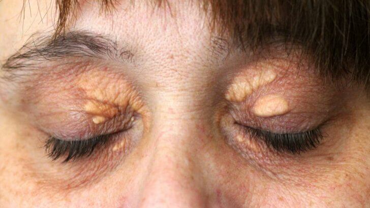
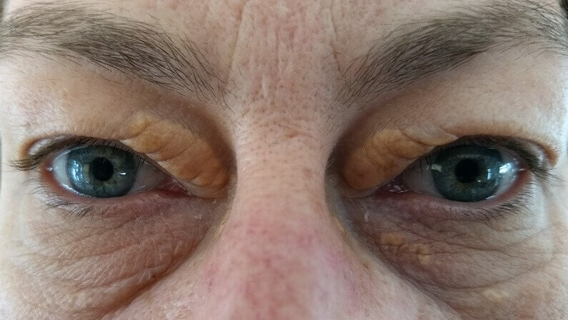
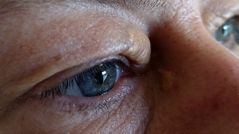
What Is A Xanthoma On The Eyelid Skin?
That’s xanthelasma, the most common skin xanthoma: soft, yellow plaques near the inner corners of the eyelids. About half of people with it have a lipid disorder, and the other half have normal blood fats.
StatPearls reports a primary or secondary lipid disorder in about half of cases, seen from about age 20 to 70 and most often between 35 and 55.[1] Secondary causes include diabetes, an underactive thyroid, primary biliary cholangitis, nephrotic syndrome and retinoid or estrogen medicines.[1] Unlike most body xanthomas, xanthelasma rarely fades even when cholesterol comes down.
Our xanthelasma guide and our page on the causes of xanthelasma cover it in full.

What Do Tendon Xanthomas Mean?
They’re a classic sign of familial hypercholesterolemia, an inherited condition that keeps LDL cholesterol high from birth. More rarely they signal cerebrotendinous xanthomatosis, a genetic condition that also affects the brain and eyes.
GeneReviews, the National Library of Medicine’s genetics reference, puts familial hypercholesterolemia at about 1 in 250 to 1 in 500 people, passed on with a 50% chance to each child, with xanthelasma and tendon xanthomas on the Achilles tendons, elbows, hands, knees and feet.[2] Without care, about half of affected men had a heart event by 50 and about 30% of women by 60; statins are the first-line medicine.[2]
Cerebrotendinous xanthomatosis is caused by changes in the CYP27A1 gene and can bring diarrhea in infancy, cataracts in about three in four children, tendon xanthomas in the teens or twenties, and nerve problems in adulthood; chenodeoxycholic acid replaces the missing bile acid, and early care appears to prevent symptoms.[6] Our pages on tendon xanthomas and Achilles tendon xanthoma go further.

What Are Tuberous And Palmar Xanthomas?
Tuberous xanthomas are firm yellow nodules over the elbows, knees and buttocks; palmar xanthomas are yellow lines in the creases of the palms and fingers. Palmar ones in particular point to dysbetalipoproteinemia, an inherited mix-up in how the body clears fats.

StatPearls describes palmar and tuberous xanthomas as features of dysbetalipoproteinemia, and notes the palmar ones fade once the condition is managed.[3]
Both are painless and slow-growing. Our pages on tuberous xanthoma and palmar xanthoma explain each in detail.

What Do Eruptive Xanthomas On The Skin Mean?
Very high triglycerides, often above 1,000 mg/dL. They appear suddenly as crops of small yellow bumps with a red rim, usually on the buttocks, arms and legs, and they need a prompt doctor’s visit because of the pancreatitis risk.
StatPearls links eruptive xanthomas with triglycerides above 1,000 mg/dL and notes that uncontrolled diabetes is a common driver; at that level the risk of pancreatitis, a painful and dangerous inflammation of the pancreas, rises too.[4] The same high triglycerides can make the retina’s blood vessels look creamy on an eye exam.[4]
Eruptive xanthomas usually settle once triglycerides come down. Our page on eruptive xanthoma explains more.

What Are Plane Xanthomas On The Skin?
Flat yellow patches that can appear on the eyelids, neck, trunk or skin folds. When they’re widespread and blood fats are normal, they can be linked with a blood protein disorder and need investigating.
Diffuse plane xanthoma with normal lipids has been linked with monoclonal gammopathy, an abnormal protein made by a group of blood cells.[5] That’s why flat yellow patches spreading beyond the eyelids, especially with normal cholesterol, call for a doctor’s check that includes blood protein tests.
Plane xanthomas in skin creases can also accompany high lipids. Our page on plane xanthoma covers both forms.

Skin Xanthoma Types Compared
| Type | Where | Look | Linked With | What To Do |
|---|---|---|---|---|
| Xanthelasma | Eyelids, inner corners | Soft, flat or slightly raised yellow plaques | A lipid disorder in about half | Blood tests; remove if you wish |
| Tendon | Achilles, knuckles, elbows, knees | Firm lumps that move with the tendon | Familial hypercholesterolemia; rarely CTX | Prompt lipid check; family testing |
| Tuberous | Elbows, knees, buttocks | Firm yellow nodules | Dysbetalipoproteinemia | Lipid check and management |
| Palmar | Palm and finger creases | Yellow lines | Dysbetalipoproteinemia | Lipid check; fades once managed |
| Eruptive | Buttocks, arms, legs | Sudden crops of small yellow-red bumps | Triglycerides above 1,000 mg/dL | See a doctor promptly |
| Plane | Eyelids, neck, trunk, folds | Flat yellow patches | High lipids, or a blood protein disorder | Doctor’s check, including blood proteins |
Which Tests Are Done For A Skin Xanthoma?
A fasting lipid profile first, then tests for causes: liver, thyroid and blood sugar, plus genetic or blood protein tests depending on the type. A biopsy is only needed when the diagnosis is uncertain.
- Examination. The type and site usually point to the likely cause.
- Fasting lipid profile. Total cholesterol, LDL, HDL and triglycerides.[1]
- Cause tests. Liver panel, thyroid function and fasting glucose, with HbA1c in diabetes.[1]
- Type-specific tests. Genetic testing for inherited lipid disorders; blood protein tests for widespread plane xanthomas.[2][5]
- Biopsy if unsure. Shows foam cells under the microscope.[1]
If an inherited condition is found, close relatives are usually offered testing too. Our page on whether xanthelasma indicates raised cholesterol explains the lipid results.

What Do Skin Xanthomas Mean For Your Health?
The deposits themselves are benign, but they’re a visible window onto your blood fats. Depending on the type, they can point to heart risk, pancreatitis risk or, rarely, a blood or genetic disorder.
- Heart risk. Tendon xanthomas mean very high LDL from birth in familial hypercholesterolemia.[2]
- Pancreatitis risk. Eruptive xanthomas mean dangerously high triglycerides.[4]
- Blood disorder. Widespread plane xanthomas with normal lipids can mean monoclonal gammopathy.[5]
- Lipid disorder. Xanthelasma comes with a lipid problem in about half of people.[1]
The good news is that most of these causes can be managed well once found. Our page on whether xanthelasma is genetic explains the family side.

Do Skin Xanthomas Go Away?
Many body xanthomas shrink or fade once the blood fat problem is controlled, especially eruptive and palmar ones. Eyelid xanthelasma is the exception: it rarely fades, even when cholesterol comes down.
StatPearls notes palmar xanthomas fade once dysbetalipoproteinemia is managed.[3] Eruptive xanthomas usually settle as triglycerides fall. Tendon xanthomas change slowly, if at all, because they sit within the tendon itself. For xanthelasma, StatPearls notes lowering lipids may do little for plaques that have already formed.[1]
That difference is why eyelid xanthelasma is usually removed if it bothers you, while body xanthomas are mostly managed through the cause.

Can Skin Xanthomas Be Removed?
Yes. Large or persistent body xanthomas can be removed surgically by a specialist after the cause is managed. Eyelid xanthelasma can be removed with laser, surgery, peels or freezing at a clinic, or at home with a cream made for xanthelasma removal.

For eyelid xanthelasma, StatPearls notes that surgery must spare enough eyelid skin to keep the lid in shape, and that plaques come back after removal in generally fewer than half of cases.[1] Body xanthomas are best removed only after the blood fat problem is under control, since they can shrink on their own once it is. Our pages on clinic options for xanthomas, removal of xanthelasma and our xanthelasma removal cream guide set out the choices.

If your xanthoma is the eyelid type, xanthelasma, and your doctor has checked the cause, this is the cream we make for removing it at home.
When Should You See A Doctor About A Skin Xanthoma?
Always, when a new one appears, for the diagnosis and blood tests. Go promptly for sudden crops of bumps, lumps on your tendons, xanthomas at a young age, or widespread flat patches.
- Sudden crops of yellow bumps. Possible very high triglycerides and pancreatitis risk.[4]
- Tendon lumps or young age. Possible inherited high cholesterol.[2]
- Widespread flat patches. Possible blood protein disorder.[5]
- Childhood cataracts or long-standing diarrhea with tendon lumps. Possible cerebrotendinous xanthomatosis.[6]
- Firm, bleeding or ulcerated spots. Rule out other skin conditions.
Our guide to what can look like xanthelasma covers the look-alikes on the eyelids.

What’s The Bottom Line On Skin Xanthomas?
Skin xanthomas are yellow deposits of fat-filled cells, and where they appear points to the cause: xanthelasma on the eyelids, tendon xanthomas with inherited high cholesterol, eruptive xanthomas with very high triglycerides, palmar xanthomas with dysbetalipoproteinemia, and widespread plane xanthomas that may signal a blood disorder. See a doctor for blood tests. Body xanthomas often fade as the cause is managed; eyelid xanthelasma usually needs removing.
Xanthel® is a cosmetic cream made for xanthelasma removal at home, sold by Xanthelasma.com. It’s for the eyelid type only, not xanthomas on the body. You’ll find it in the removal section above and at the foot of this page, or read more about Xanthel® for eyelid xanthoma removal. For the full family, see our xanthomas guide, or browse all our xanthelasma guides.
Common Questions About Skin Xanthomas
What Is A Skin Xanthoma?
A yellow deposit in the skin made of immune cells packed with fat, often a sign of a blood fat problem.
What Are The Types Of Skin Xanthoma?
Xanthelasma on the eyelids, plus tendon, tuberous, palmar, eruptive and plane xanthomas.
Are Skin Xanthomas Dangerous?
The deposits are benign, but they can signal high cholesterol, very high triglycerides or, rarely, a blood or genetic disorder.
Do Skin Xanthomas Mean I Have High Cholesterol?
Often, but not always; about half of people with eyelid xanthelasma have normal blood fats, and some types point to triglycerides instead.
What Causes Sudden Yellow Bumps On The Skin?
Crops of small yellow bumps can be eruptive xanthomas from very high triglycerides, which need a prompt doctor’s visit.
Do Skin Xanthomas Go Away?
Many body xanthomas shrink once blood fats are controlled, but eyelid xanthelasma rarely fades by itself.
Can Skin Xanthomas Be Removed?
Yes; body xanthomas can be removed surgically, and eyelid xanthelasma by laser, surgery or a cream made for xanthelasma removal at home.
Should I See A Doctor About A Skin Xanthoma?
Yes, every new xanthoma deserves a doctor’s check and blood tests, and promptly if it appeared suddenly or is on a tendon.
Sources
- Blair K. Xanthelasma Palpebrarum. StatPearls, National Library of Medicine (NCBI Bookshelf), updated 2026.
- Ison HE, Clarke SL, Knowles JW. Familial Hypercholesterolemia. GeneReviews, National Library of Medicine (NCBI Bookshelf), revised 2025.
- Javvaji A, Can AS, Sharma S. Dysbetalipoproteinemia. StatPearls, National Library of Medicine (NCBI Bookshelf), updated 2024.
- Mishra C, Tripathy K. Lipemia Retinalis. StatPearls, National Library of Medicine (NCBI Bookshelf), updated 2026.
- Szalat R, et al. Pathogenesis and treatment of xanthomatosis associated with monoclonal gammopathy. Blood, 2011.
- Federico A, Gallus GN. Cerebrotendinous Xanthomatosis. GeneReviews, National Library of Medicine (NCBI Bookshelf), revised 2024.