
What A Yellow Spot In Your Stomach Means, How It’s Linked To Gastritis, And Why Doctors Keep An Eye On It
A gastric xanthoma is a small, harmless yellow-white patch of fat-filled immune cells in the stomach lining, usually found by chance during an endoscopy. The spot itself doesn’t need removing and doesn’t turn into cancer. What matters is the stomach lining around it, because gastric xanthomas often appear alongside gastritis, stomach lining thinning and H. pylori infection. Here’s what xanthoma gastric findings mean, what causes them and what happens next.
Gastric xanthoma is a small, benign yellow-white spot of fat-filled immune cells in the stomach lining, usually 1 to 10 mm and found by chance on endoscopy. It needs no removal, but it’s often linked to H. pylori, atrophic gastritis and intestinal metaplasia, so doctors check the rest of the stomach carefully.
Key Takeaways
- Gastric xanthoma is a small, benign yellow-white spot in the stomach lining.
- It’s found in roughly 2% to 7% of people having an endoscopy.
- It’s made of immune cells filled with fat, not of cancer cells.
- It’s linked with H. pylori, atrophic gastritis and intestinal metaplasia.
- Some studies suggest it may flag a higher stomach cancer risk, so follow-up matters.
- It isn’t the same as eyelid xanthelasma, which is a skin condition.
On This Page 14 sections
What Is Gastric Xanthoma?
Gastric xanthoma, also called gastric xanthelasma, is a small, benign collection of fat-filled immune cells, known as foam cells, in the stomach lining. It shows up on endoscopy as a yellow or white spot and is almost always harmless in itself.
Xanthomas are deposits of fat-filled cells, and they can form in many parts of the body: the eyelids, the skin, the tendons and, less obviously, the lining of the gut. When one forms in the stomach, doctors call it a gastric xanthoma. You may also see it written as gastric xanthelasma or gastric xanthomatosis when there are several. Our guide to xanthomas covers the whole family.
Under the microscope, a gastric xanthoma is a cluster of foamy histiocytes, a type of macrophage, packed with fat. These cells sit in the lamina propria, the thin layer of tissue just under the stomach’s surface.[5] Researchers think they form as part of the stomach’s response to damage or ageing of its lining.[3] Because they’re made of immune cells rather than stomach lining cells, they aren’t a tumor and don’t grow into one.
Gastric xanthoma was first described in 1929, and for decades it was seen as a curiosity of little importance.[4] More recent research has changed that view, not because the spot itself is dangerous, but because of what it tends to sit alongside. For a shorter introduction, see our page on what gastric xanthelasma is.

What Does Gastric Xanthoma Look Like, And Where Does It Form?
On endoscopy it looks like a small, flat or slightly raised, well-defined yellow-white plaque, usually 1 to 10 mm across. It’s most often found in the lower part of the stomach, the antrum, though it can appear in the body and upper stomach too.
Endoscopists describe gastric xanthomas as yellow-white plaques that are flat or just slightly raised, typically between 1 and 10 mm in size.[1] In one Korean study of 54 people with the finding, the average size was 7 mm, none was bigger than 10 mm, and most people (64.8%) had a single spot.[4] The same study found them in these parts of the stomach:
- The antrum, the lower stomach near the outlet: 53.7%.
- The body, the large middle part: 35.2%.
- The fundus, the dome at the top: 11.1%.[4]
They can rarely appear in other parts of the gut, such as the esophagus or the first part of the small intestine (the duodenum), but these are much less common than in the stomach.[1][5] Because they’re so small and pale, they’re easy to overlook, and finding them partly depends on how carefully the endoscopist is looking.

How Common Is Gastric Xanthoma?
It’s a fairly common chance finding. Large endoscopy studies report it in about 2% to 7% of patients, and it’s more common with age. It affects men and women about equally in most studies.
Figures vary by country and by study, partly because rates of stomach conditions differ around the world:
- A Chinese study of 69,071 people having upper endoscopy found gastric xanthoma in 1.77%.[1]
- A Japanese health-checkup study of 1,823 people found it in 5.9%.[2]
- A Korean study of 771 endoscopies found it in 7%.[4]
Age is the clearest pattern. In the large Chinese study, people with gastric xanthoma were older on average (53 against 46), and the odds rose with each year of age.[1] Sex made no difference in that study or the Korean one,[1][4] although the Japanese study found it more often in men and in people aged 65 or over.[2] Gastric xanthoma is rarely seen in young people, and finding one is usually just a sign of a stomach lining that has been through some wear and tear.

What Causes Gastric Xanthoma?
The exact cause isn’t settled, but the leading idea is that immune cells clean up fat-rich debris from a damaged or inflamed stomach lining, fill up with it and become foam cells. Long-term inflammation, H. pylori infection and ageing of the lining all seem to play a part.
Your stomach lining is constantly being worn down and repaired, especially when it’s inflamed. When lining cells are damaged, their fat-rich membranes become debris that macrophages swallow. When those macrophages take in more fat than they can break down, they become foam cells, and a cluster of them forms the yellow spot.[5] Researchers also think free radicals released during inflammation may contribute.[3]
Several triggers have been proposed:
- Chronic inflammation (gastritis). The strongest and most consistent link across studies.[1][4]
- H. pylori infection. One theory is that macrophages become foamy after swallowing the bacteria, and H. pylori material has been found in foam cells.[5]
- Ageing of the stomach lining. Gastric xanthoma becomes more common with every decade.[1]
- Blood fats. One study found higher LDL and lower HDL cholesterol in people with gastric xanthoma, though the role of blood fats is still unclear.[4]
- Diabetes. Linked with gastric xanthoma in the Japanese checkup study.[2]
This is a different process from eyelid xanthelasma, where fats leak out of eyelid blood vessels into the skin. If you’re curious about that one, our guide to the causes of xanthelasma explains it.

Is Gastric Xanthoma Linked To H. Pylori And Gastritis?

Yes. Gastric xanthoma is strongly linked with atrophic gastritis (thinning of the stomach lining) and intestinal metaplasia (lining that has changed to look like intestine), and more weakly with H. pylori infection. That’s why it’s seen as a warning sign to check the rest of the stomach.
The largest study so far, of over 69,000 endoscopies, found that people with gastric xanthoma were far more likely to have these changes than people without it:[1]
- Intestinal metaplasia in 51.9%, against 19.3%.
- Atrophic gastritis in 42.9%, against 14.3%.
- H. pylori infection in 34.6%, against 27.0%.
After allowing for age and other factors, intestinal metaplasia more than doubled the odds of gastric xanthoma, atrophic gastritis raised them by about 80%, and H. pylori by about a third. People who had H. pylori together with intestinal metaplasia had the highest rate, at about 5%. People with several xanthomas were more likely still to have atrophy and metaplasia.[1] The authors’ conclusion was simple: endoscopists should take extra time over patients with gastric xanthoma, especially multiple ones.[1]
The Korean study also found atrophic gastritis three times as often in people with gastric xanthoma (44.4% against 14.8%), though in a population where H. pylori was very common it found no difference in infection rates.[4] One more point: clearing H. pylori with antibiotics doesn’t seem to make an existing xanthoma disappear.[3]

Is Gastric Xanthoma A Sign Of Stomach Cancer?
The xanthoma itself isn’t cancer and doesn’t become cancer. Some studies, mainly from Japan, suggest it may be a marker of a stomach lining at higher risk, while the largest study found no independent link once gastritis changes were taken into account. Either way, it’s a reason for careful follow-up, not alarm.
The research is worth knowing in proportion. In a Japanese study of 1,823 people having health checkups, early stomach cancer later developed in 14.0% of those with gastric xanthoma, against 0.8% of those without. After allowing for other factors, gastric xanthoma was linked with almost six times the odds of developing early gastric cancer, alongside extensive atrophy of the lining.[2] A second Japanese study followed 115 people who’d had an early stomach cancer removed endoscopically, for a median of 55 months. New cancers developed in 27.1% of those with gastric xanthoma, against 8.9% without, and xanthoma was an independent predictor.[3]
The large Chinese study adds balance. Gastric cancer was slightly more common in people with xanthoma (1.5% against 0.9%), but once atrophy, metaplasia and H. pylori were allowed for, xanthoma wasn’t an independent risk factor.[1] In other words, the xanthoma may mainly be a visible sign of the inflamed, thinned lining that carries the risk.
What this means for you: most people with a gastric xanthoma never develop stomach cancer. But it’s sensible for your gastroenterologist to look closely at the rest of the stomach, take biopsies where needed, and decide whether follow-up endoscopies make sense for you. This is very different from eyelid xanthelasma, which isn’t linked with cancer at all, as our guide on whether xanthelasma can be cancerous explains.

Does Gastric Xanthoma Cause Symptoms?
No. Gastric xanthoma itself is silent. Any stomach symptoms you have, such as indigestion or pain, come from whatever led to the endoscopy, often gastritis or another condition, not from the xanthoma.
Because the spots are tiny and sit just under the surface, they don’t cause pain, bleeding or blockage. That’s why almost every gastric xanthoma is found by chance, during an endoscopy done to look into heartburn, indigestion, anemia, suspected H. pylori or a routine check. If you have ongoing symptoms after being told you have one, raise them with your doctor, since the cause lies elsewhere.
Some symptoms always deserve a prompt check whatever was found before:
See Your Doctor Promptly If
- You’re losing weight without trying
- Swallowing is difficult or painful
- You vomit blood or pass black stools
- Indigestion is new and persistent in later life
Usually Routine If
- The xanthoma was a chance finding
- Biopsies were reassuring
- You have a follow-up plan
- Any H. pylori has been dealt with

How Is Gastric Xanthoma Diagnosed?
It’s spotted during an upper endoscopy and confirmed by a small biopsy, which shows fat-filled macrophages. Special stains help the pathologist tell it apart from lookalikes, including a rare type of stomach cancer.
The process usually goes like this:
- Upper endoscopy. A thin camera is passed into the stomach, often for an unrelated reason, and the endoscopist sees a small yellow-white plaque.
- Biopsy. A tiny sample is taken painlessly through the scope.
- Microscope and stains. The pathologist sees clusters of foamy cells. They stain positive for CD68, a macrophage marker, and negative for mucin and epithelial markers, confirming they’re immune cells.[5]
- Ruling out lookalikes. These stains matter because xanthoma cells can resemble signet ring cell carcinoma, a type of stomach cancer, under the microscope.[5]
- Checking the surrounding lining. Biopsies elsewhere in the stomach look for H. pylori, atrophic gastritis and intestinal metaplasia.[1]
Your report may simply say “gastric xanthoma” or “xanthelasma of the stomach”. If it also mentions atrophy or metaplasia, ask your doctor what that means for follow-up. For more on the terms doctors use, see our xanthoma definition page.

Does Gastric Xanthoma Mean You Have High Cholesterol?
Not necessarily. Unlike some skin xanthomas, gastric xanthoma isn’t a reliable sign of high cholesterol. One study found slightly higher LDL and lower HDL in people with it, but total cholesterol and triglycerides were no different.
It’s a reasonable question, since all xanthomas contain fat. In the Korean study, people with gastric xanthoma had higher LDL (about 113 against 96 mg/dL) and lower HDL (about 49 against 63 mg/dL) than matched people without it, while total cholesterol, triglycerides and body weight didn’t differ.[4] The author was clear that it’s not known whether blood fats actually cause gastric xanthoma.[4]

So a gastric xanthoma alone isn’t a reason to assume you have a cholesterol problem, but a routine cholesterol check is never a bad idea, especially in midlife. Eyelid xanthelasma is a different story: about half of people with it have a blood fat disorder.[6] Our page on whether xanthelasma indicates raised cholesterol explains that link.

What Happens After Gastric Xanthoma Is Found?
The xanthoma itself is left alone. Care focuses on the stomach lining: clearing any H. pylori, assessing any atrophy or metaplasia, and deciding with your gastroenterologist whether and when to repeat the endoscopy.
Because the xanthoma is benign and silent, there’s nothing to gain from removing it. Instead, your doctor will look at the bigger picture:
- H. pylori. If it’s found, a course of antibiotics and acid-reducing medicine is usually offered to clear it, then a test to confirm it’s gone.
- Atrophy and metaplasia. If biopsies show these changes, your doctor will assess how extensive they are.
- Surveillance. Depending on the findings, you may be offered repeat endoscopies at set intervals. People with several xanthomas, extensive atrophy or a previous stomach cancer are the ones doctors watch most closely.[1][2][3]
- General health. Diabetes control, a cholesterol check and not smoking all support a healthy stomach and heart.
This is medical care led by your doctor. There’s no cream, diet or home remedy for a gastric xanthoma, and none is needed.
Gastric Xanthoma Compared With Other Xanthomas
| Type | Where | How It’s Found | Main Links | What Happens Next |
|---|---|---|---|---|
| Gastric xanthoma | Stomach lining | By chance on endoscopy | Gastritis, atrophy, metaplasia, H. pylori | Left alone; the stomach lining is checked and followed up |
| Xanthelasma | Eyelid skin | Seen in the mirror | Blood fat disorders in about half | Cholesterol check, then removal if wanted |
| Tendon xanthomas | Achilles, hands, elbows, knees | Firm lumps on tendons | Inherited high cholesterol | Medical care for blood fats |
| Eruptive xanthomas | Skin of buttocks, arms, legs | Sudden crops of small bumps | Very high triglycerides | Urgent blood fat assessment |
How Is Gastric Xanthoma Different From Eyelid Xanthelasma?
They share a name and the same foam cells, but they’re different conditions. Gastric xanthoma is an internal finding tied to the stomach lining; eyelid xanthelasma is a visible skin condition tied in about half of people to blood fats, and it can be removed.
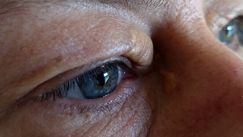
Eyelid xanthelasma is the soft yellow plaques near the inner corners of the eyes. It affects about 1.1% of women and 0.3% of men, and it forms when fats leak from eyelid blood vessels and collect in skin cells.[6] Having a gastric xanthoma doesn’t mean you’ll get eyelid plaques, or the other way round. Our guides to xanthelasma and to what causes xanthelasma cover the eyelid form, and is xanthelasma dangerous puts its health links in proportion.
The practical difference is what you can do about it. A gastric xanthoma is left alone. Eyelid xanthelasma rarely fades by itself, and once a doctor has checked your blood fats, it can be removed at a clinic or at home. Our guide to the removal of xanthelasma covers the options.
What’s The Bottom Line On Gastric Xanthoma?
Gastric xanthoma is a small, benign yellow spot of fat-filled immune cells in the stomach lining. It needs no removal, but it often sits alongside gastritis, atrophy and metaplasia, so it’s a cue for a careful look at the stomach and a sensible follow-up plan.
If you’ve been told you have one, ask your gastroenterologist three things: was H. pylori found, did the biopsies show atrophy or metaplasia, and do you need a repeat endoscopy. For more on the whole family, see our guides to xanthoma and xanthomatosis. If you also have yellow plaques on your eyelids, that’s a separate skin matter, and our xanthelasma guide is the place to start. You can browse all our xanthelasma guides too.
Common Questions About Gastric Xanthoma
Is Gastric Xanthoma Dangerous?
No, the xanthoma itself is benign and doesn’t turn into cancer. It’s often found alongside gastritis and changes in the stomach lining, though, so your doctor may want to follow those up.
Is Gastric Xanthoma Cancer?
No. It’s made of fat-filled immune cells, not cancer cells. Some studies link it with a higher risk of stomach cancer elsewhere in the lining, which is why careful follow-up is advised.
Does Gastric Xanthoma Need To Be Removed?
No. It causes no symptoms and is harmless in itself, so it’s left alone. Care focuses on the surrounding stomach lining instead.
Is Gastric Xanthoma Caused By H. Pylori?
It may be one trigger. Gastric xanthoma is more common in people with H. pylori, and even more so with atrophic gastritis or intestinal metaplasia, but the exact cause isn’t settled.
Will Gastric Xanthoma Go Away After H. Pylori Is Cleared?
Usually not. Research suggests gastric xanthomas tend to persist after H. pylori has been cleared, which isn’t a concern in itself.
Does Gastric Xanthoma Mean I Have High Cholesterol?
Not necessarily. One study found slightly higher LDL and lower HDL in people with it, but it isn’t a reliable sign of high cholesterol, and a routine check is enough.
Do I Need Another Endoscopy?
That depends on what else was found. People with several xanthomas, extensive atrophy, intestinal metaplasia or a past stomach cancer are more likely to be offered follow-up endoscopies.
Is Gastric Xanthoma The Same As Xanthelasma On The Eyelids?

No. Both contain fat-filled cells, but gastric xanthoma is an internal stomach finding, while eyelid xanthelasma is a skin condition that can be removed once a doctor has checked your blood fats.
Sources
- Feng L, Zhang M, Guan J, et al. Gastric xanthelasma is a warning sign for Helicobacter pylori infection, atrophic gastritis, and intestinal metaplasia. Frontiers in Medicine, 2023;10:1252346.
- Sekikawa A, Fukui H, Sada R, et al. Gastric atrophy and xanthelasma are markers for predicting the development of early gastric cancer. Journal of Gastroenterology, 2016;51:35-42.
- Fukushima M, Fukui H, Watari J, et al. Gastric Xanthelasma, Microsatellite Instability and Methylation of Tumor Suppressor Genes in the Gastric Mucosa: Correlation and Comparison as a Predictive Marker for the Development of Synchronous/Metachronous Gastric Cancer. Journal of Clinical Medicine, 2022;11(1):9.
- Yi SY. Dyslipidemia and H pylori in gastric xanthomatosis. World Journal of Gastroenterology, 2007;13(34):4598-4601.
- Andrejic BM, Bozanic SV, Solajic NS, Djolai MA, Levakov AM. Xanthomas of the stomach: a report of two cases. Bosnian Journal of Basic Medical Sciences, 2012;12(2):127-129.
- Blair K. Xanthelasma Palpebrarum. StatPearls, National Library of Medicine (NCBI Bookshelf), updated 2026.