The First Signs, Where It Begins, What Sets It Off Under The Skin, And What To Do When You Spot Your First Yellow Patch
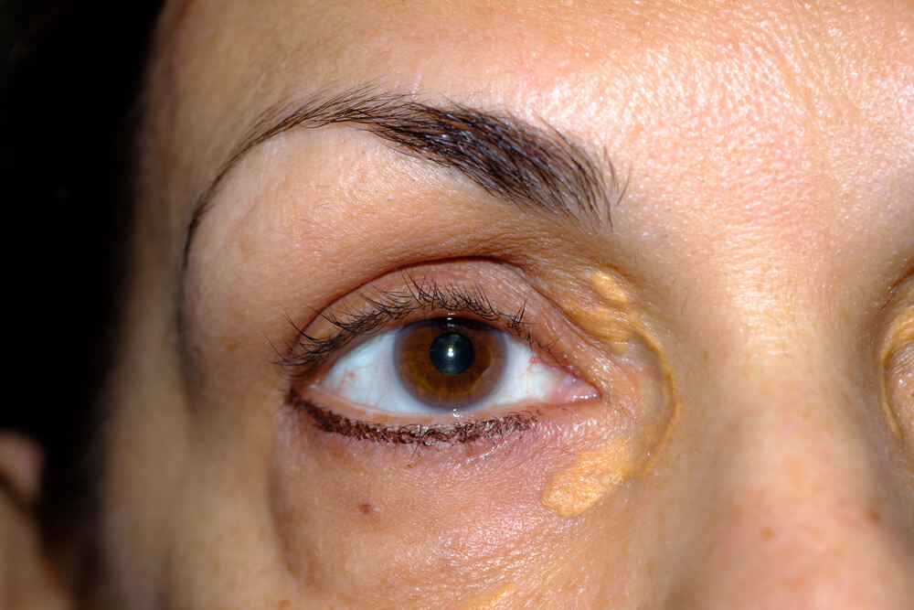
Xanthelasma usually starts as a small, soft, pale yellow patch near the inner corner of an eyelid, most often the upper lid. It’s painless, doesn’t itch and is easy to mistake for a blemish at first. Underneath, fat-filled immune cells are gathering in the eyelid skin. This guide explains what the first signs look like, why it starts, who it starts in, and what to do when you notice one.
Xanthelasma starts when fats leak from tiny eyelid blood vessels and are swallowed by immune cells, which turn into fat-filled foam cells and gather in the skin. You first see a small, soft, flat or slightly raised yellow patch near the inner corner of the eye, often on both sides. It usually stays the same or grows slowly, so it’s worth getting your blood fats checked and acting while the patch is small.
Key Takeaways
- It starts as a small, soft, pale yellow patch near the inner corner of the eye.
- The upper lid is the most common starting point, often on both sides.
- It begins when fat-filled immune cells gather in the eyelid skin.
- It most often starts between 35 and 55, and in women more than men.
- Many people have normal cholesterol when it starts.
- Small, early plaques are the easiest to remove.
On This Page 18 sections
How Does Xanthelasma Start?
It starts as a small, soft, pale yellow patch in the eyelid skin, usually near the inner corner of the eye. Beneath it, fat-filled immune cells called foam cells are collecting around tiny blood vessels.
Xanthelasma palpebrarum is the most common form of xanthoma, a deposit of fat-laden immune cells in the skin.[1] It doesn’t appear overnight. The process under the skin builds up quietly, and by the time you notice a faint yellow mark, the foam cells have already gathered in enough numbers to show through the thin eyelid skin.
For the condition as a whole, see our main xanthelasma guide. Related pages cover how xanthelasma forms and how you get xanthelasma; this one focuses on the very beginning.
What Do The First Signs Of Xanthelasma Look Like?
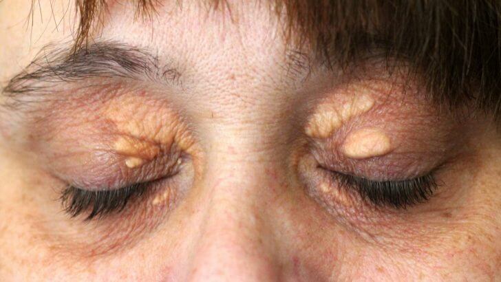
A small, flat or slightly raised, soft patch that’s pale yellow or cream colored. It’s painless, doesn’t itch, and may look like dry skin or a blemish at first.
- Color. Pale yellow to cream, sometimes faint enough to miss in dim light.
- Feel. Soft and smooth, not hard or gritty.
- Shape. A small oval or irregular patch, often only a few millimeters across.
- Height. Flat or just slightly raised.
- Sensation. No pain, itching, discharge or redness.
- Pattern. Often on both sides, in matching positions.[2]
Many people first notice it in a photo or under bright bathroom light. If a spot is red, sore, crusting or grew within days, it’s more likely to be something else. Our guide on what xanthelasma looks like shows the typical appearance at different stages.

Where On The Eyelid Does Xanthelasma Start?
Near the inner corners of the eyes, close to the nose. It starts more often on the upper lid than the lower lid, and frequently appears on both sides.
The American Academy of Ophthalmology’s EyeWiki describes xanthelasma as usually appearing near the inner corners of the eyelids, often on both sides and more often on the upper lid.[2] Lower-lid plaques do occur, sometimes later or alongside upper-lid ones. Starting on one side and then appearing on the other is common, so a matching patch a few months later isn’t unusual. If you’re noticing yellow marks under the eyes specifically, our guide on xanthelasma under the eyes covers the lower lid.

What’s Happening Under The Skin When Xanthelasma Starts?
Fats leak from tiny eyelid blood vessels, immune cells swallow them and turn into foam cells, and those cells gather around the vessels with some inflammation. That cluster is what you see as the first yellow patch.
- Constant blinking. Blinking repeatedly squeezes the eyelid’s tiny capillaries, and with age they become leakier.[1]
- Fats seep out. Cholesterol-carrying particles leak into the surrounding skin.[1]
- Immune cells respond. Macrophages swallow the fats to clear them.[1]
- Foam cells form. Overloaded macrophages become foam cells, with some inflammation.[1]
- A patch appears. As foam cells collect, the yellow color shows through the thin eyelid skin.
That mechanical stress is the leading explanation for why xanthelasma favors the eyelids, and it helps explain why plaques often appear in matching positions on both sides.[1] Our guide on why xanthelasma occurs explains the process in more depth.

Who Does Xanthelasma Start In, And At What Age?
It affects about 1.1% of women and 0.3% of men, and usually starts between 20 and 70, most often between 35 and 55.
Those figures come from StatPearls.[1] In a study of 42 patients, 85.7% were women and the average age was about 47, and the authors listed hormonal factors among the possible explanations.[5] The age pattern also fits the mechanism, since eyelid vessels become leakier over time. So the most typical picture is a woman in midlife noticing a faint yellow patch, but it can start in men and at other ages too. Our guide on whether xanthelasma is common has more on who gets it.
What Does It Mean If Xanthelasma Starts When You’re Young?
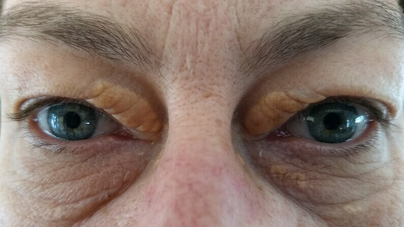
An inherited blood fat disorder is more likely. If it starts in your twenties or earlier, a full lipid profile is especially important.
StatPearls advises considering an inherited blood fat disorder when xanthelasma appears in young people.[1] One example is familial hypercholesterolemia, which causes very high LDL cholesterol from birth; its signs include xanthelasma and lumps on the tendons, and statins are the first-line medicine.[3] Finding it early matters because it raises heart risk from a young age, and close relatives may have it too. Our guide on whether xanthelasma is genetic explains the inherited side.

What Sets Off Xanthelasma In The First Place?
Blood fat disorders are the main known trigger, but between 25% and 70% of people have normal blood fats when it starts. Some health conditions and medicines can also play a part.

StatPearls lists primary lipid disorders, including types IIa, IIb and IV hyperlipidemia and low HDL, and secondary causes such as diabetes, an underactive thyroid, primary biliary cholangitis, nephrotic syndrome and retinoid or estrogen medicines.[1] A meta-analysis of 15 studies and 854 patients found higher total cholesterol, LDL and apolipoprotein B, and lower apolipoprotein A1, in people with xanthelasma.[4]
Yet many people have normal results. EyeWiki gives the 25% to 70% range for normal lipids.[2] In the study of 42 patients, 54.8% had normal cholesterol, but their HDL was markedly lower than in people without xanthelasma, and the authors suggested local, hormonal or immune cell factors.[5] So a normal cholesterol number doesn’t always mean a normal lipid profile. Our guide to the causes of xanthelasma covers each one.

What Can Early Xanthelasma Be Confused With?
Early patches are sometimes mistaken for dry skin, a blemish, milia, syringoma or sebaceous hyperplasia. A doctor can usually tell the difference by looking.
EyeWiki lists syringoma and sebaceous hyperplasia among the lookalikes, along with a few rarer conditions that may need a biopsy.[2] Styes and inflamed bumps are red and tender and settle within days or weeks, while xanthelasma is yellow, soft, painless and persistent. If a spot is hard, bleeds, ulcerates or changes quickly, see a doctor promptly rather than assuming it’s xanthelasma. Our guide on what can be mistaken for xanthelasma sets out the differences.

How Xanthelasma Typically Develops After It Starts
| Stage | What You See | Typical Timing | What To Do |
|---|---|---|---|
| Just starting | Faint, flat, pale yellow patch near the inner corner | First noticed | See a doctor and get blood tests |
| Established | Clearer yellow plaque, soft, sometimes slightly raised | Months | Photograph it; consider early removal |
| Spreading | Matching patch on the other side or a new one nearby | Months to years | Manage any cause; review removal options |
| Larger or merged | Thicker plaques that may join together | Years | Clinic removal with an eyelid specialist |
| Small to medium, any stage | Typical plaque, confirmed by a doctor | Whenever you choose | Xanthel® at home: one application per plaque |
How Does Xanthelasma Develop Once It Has Started?
It usually stays the same or slowly gets larger, rather than fading. New patches may appear nearby or on the other eyelid, and neighboring plaques can join together.
StatPearls notes that xanthelasma tends to stay the same or slowly enlarge once it has formed.[1] The pace varies a lot between people: some plaques barely change for years, while others grow over months. Deeper plaques matter, because plaques more than about 1 mm deep come back more often after removal.[1] Our guides on how fast xanthelasma grows and whether it can get bigger describe the typical timeline.

Can You Stop Xanthelasma Once It Has Started?
You can’t usually make an existing patch fade by lifestyle alone, but managing any cause may slow things down and lower the chance of new plaques. Removing the patch is what makes it go.
StatPearls says lifestyle changes and lipid-lowering medicine may have only a limited effect on how established plaques look.[1] Xanthelasma rarely goes away without help; see whether it can go away on its own or just disappear. Getting blood fats, blood sugar and thyroid under control is still worthwhile, mainly for your heart, and may help stop the process that started the plaque from continuing.

What Should You Do When You Spot Your First Xanthelasma Patch?
See your doctor to confirm it and check your blood fats, photograph the patch, avoid squeezing or home remedies, and decide whether to remove it while it’s small.
- Book a doctor’s appointment. To confirm it’s xanthelasma and rule out lookalikes.
- Ask for blood tests. A fasting lipid profile, liver panel, thyroid tests and fasting glucose are the usual checks.[1]
- Photograph it. Daylight, no makeup, same angle, so you can track any change.
- Leave it alone. Don’t squeeze, pick or apply garlic or vinegar; see whether you can squeeze out xanthelasma.
- Manage any cause found. Diet, activity, not smoking and any medicine your doctor recommends.
- Decide on removal. Small, shallow plaques are the easiest to remove.
Our guide on what xanthelasma indicates explains what the tests look for.

Is The Start Of Xanthelasma A Warning Sign?
It can be a useful clue to check your heart health. Xanthelasma is linked with higher heart risk, even in people with normal cholesterol, though most people with it won’t have a heart attack.
A study that followed almost 13,000 people from the general population for decades found that those with xanthelasma had a higher risk of heart attack and heart disease, independent of cholesterol.[6] The meta-analysis of 854 patients also found thicker carotid artery walls and a higher risk of atherosclerosis in people with xanthelasma.[4] That’s why the first patch is a good prompt to see your doctor, who can assess your overall risk using your blood pressure, blood fats, blood sugar, smoking and family history.
Is Xanthelasma Easier To Remove When It Has Just Started?
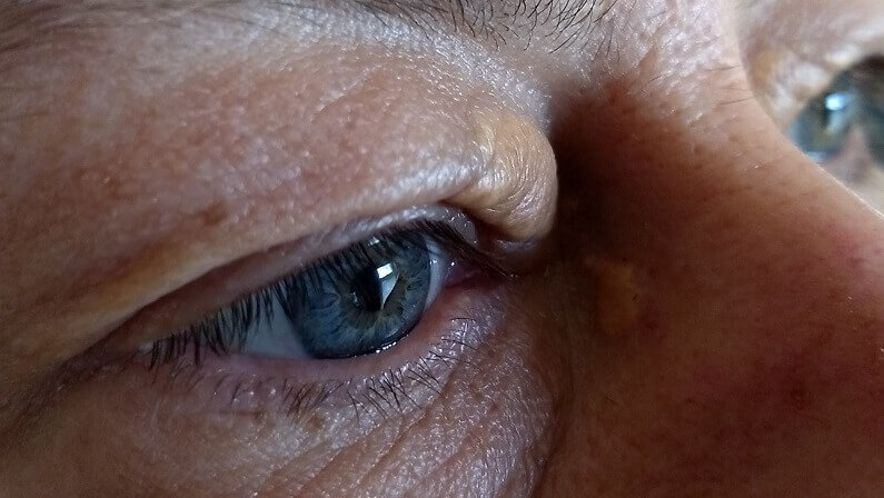
Generally, yes. Small, shallow plaques need fewer sessions, heal more quickly and come back less often than deep or large ones.
Deeper plaques are the most likely to return after removal.[1] In a 2024 acid peel study, 49.4% of patients needed just one session and 40.3% two, with plaques improved in 90.9% overall.[7] In an older long-term study of bichloracetic acid, 72% of plaques needed no further removal over an average of 68 months.[8] Clinic options include laser, acid peels, radiofrequency and surgery, while small to medium, typical plaques can also be removed at home with a cream made for xanthelasma. Our guides on how xanthelasma is removed and xanthelasma cream compare the routes.
If you’ve spotted an early patch and your doctor has checked the cause, this is the cream we make for removing xanthelasma at home.

How Can You Cover A Xanthelasma Patch That Has Just Started?
A small dab of lavender or peach color corrector followed by a thin layer of skin-tone concealer hides most early patches well, because they’re usually flat and faint.
Early patches are often easier to cover than established ones, since there’s little raised texture to show through. Use products made for the eye area, apply with a clean fingertip or small brush, and set lightly with powder so it lasts. Makeup hides the patch but doesn’t stop it growing, so it works best alongside a doctor’s check. Our guide on how to cover xanthelasma explains the routine step by step.

Can You Stop New Xanthelasma Patches From Starting?
Not always, because age, sex and inherited traits can’t be changed. But keeping blood fats, blood sugar and thyroid under control, eating well, staying active and not smoking lowers the chance of new plaques.
In the long-term bichloracetic acid study, most recurrences were in people with high cholesterol,[8] which suggests that managing blood fats matters for keeping new patches away. StatPearls puts recurrence after removal at under 50% overall, higher for deep plaques.[1] Our guides on how to prevent xanthelasma and whether xanthelasma can come back explain what helps most.

What’s The Bottom Line On How Xanthelasma Starts?
Xanthelasma starts as a small, soft, pale yellow patch near the inner corner of the eye, as fat-filled immune cells gather in the eyelid skin. See your doctor when you spot the first one, manage any cause, and remove it while it’s small if you want it gone.
Xanthel® is a cosmetic cream made for xanthelasma removal at home, sold by Xanthelasma.com. You’ll find it in the box above, or read more about Xanthel® xanthelasma removal cream, and browse all our xanthelasma guides.
Common Questions About How Xanthelasma Starts
How Does Xanthelasma Start?
As a small, soft, pale yellow patch near the inner corner of the eye, as fat-filled immune cells gather in the eyelid skin.
What Are The First Signs Of Xanthelasma?
A flat or slightly raised, soft, pale yellow patch that’s painless and doesn’t itch, often on both sides near the nose.
Where Does Xanthelasma Usually Start?
Near the inner corners of the eyes, more often on the upper lid, and frequently on both sides.
At What Age Does Xanthelasma Start?
Usually between 20 and 70, most often between 35 and 55. If it starts younger, an inherited blood fat disorder is more likely.
Does Xanthelasma Start Because Of High Cholesterol?
Sometimes, but between 25% and 70% of people have normal blood fats when it starts. Low HDL, hormones and local factors may also play a part.
How Fast Does Xanthelasma Develop After It Starts?
It usually stays the same or grows slowly over months to years, and new patches may appear nearby or on the other side.
What Should I Do When Xanthelasma Starts?
See your doctor for a check and blood tests, photograph the patch, avoid squeezing or home remedies, and consider removing it while it’s small.
Is Xanthelasma Easier To Remove When It First Starts?
Generally yes. Small, shallow plaques need fewer sessions and come back less often than deep or large ones.
Sources
- Blair K. Xanthelasma Palpebrarum. StatPearls, National Library of Medicine (NCBI Bookshelf), updated 2026.
- Patadia A, Rana HS, Stewart K, Yen MT, Lee H. Xanthelasma. EyeWiki, American Academy of Ophthalmology, reviewed September 2026.
- Ison HE, Clarke SL, Knowles JW. Familial Hypercholesterolemia. GeneReviews, National Library of Medicine (NCBI Bookshelf), revised 2025.
- Chang HC, Sung CW, Lin MH. Serum lipids and risk of atherosclerosis in xanthelasma palpebrarum: a systematic review and meta-analysis. Journal of the American Academy of Dermatology, 2020;82(3):596-605.
- Kavoussi H, Ebrahimi A, Rezaei M, Ramezani M, Najafi B, Kavoussi R. Serum lipid profile and clinical characteristics of patients with xanthelasma palpebrarum. Anais Brasileiros de Dermatologia, 2016;91(4):468-471.
- Christoffersen M, Frikke-Schmidt R, Schnohr P, Jensen GB, Nordestgaard BG, Tybjaerg-Hansen A. Xanthelasmata, arcus corneae, and ischaemic vascular disease and death in general population: prospective cohort study. BMJ, 2011.
- Sapra S, Tran JVH, Gurm H, Eleuterio M. Treatment of Xanthelasma Palpebrarum Using Trichloroacetic Acid 80%. Journal of Clinical and Aesthetic Dermatology, 2024;17(11):17-22.
- Haygood LJ, Bennett JD, Brodell RT. Treatment of xanthelasma palpebrarum with bichloracetic acid. Dermatologic Surgery, 1998;24(9):1027-1031.