What The Yellow Eyelid Plaques Are Made Of, How They Form, What They Mean For You, And How To Get Rid Of Them
Xanthelasma: what are they? They’re soft, yellow plaques on the eyelids made of immune cells that have filled up with fat, usually near the inner corners of the eyes. They’re harmless and don’t affect your vision, but they can be a sign worth a cholesterol check, and they rarely go away by themselves. Here’s what they are, how they form, and what you can do about them.
Xanthelasma are soft yellow plaques on the eyelids, made of fat-filled immune cells called foam cells. They form when fat leaks from tiny eyelid blood vessels. They affect about 1.1% of women and 0.3% of men, are harmless and painless, and are linked to a blood fat disorder in about half of people. They can be removed.
Key Takeaways
- Xanthelasma are fat-filled immune cells collected in the eyelid skin.
- They usually appear near the inner corners of the upper eyelids.
- They’re not cancer, not contagious and don’t affect your vision.
- About half of people with them have a blood fat disorder.
- They’ve been linked with a higher risk of heart disease.
- They rarely fade on their own, but they can be removed.
On This Page 16 sections
Xanthelasma: What Are They?
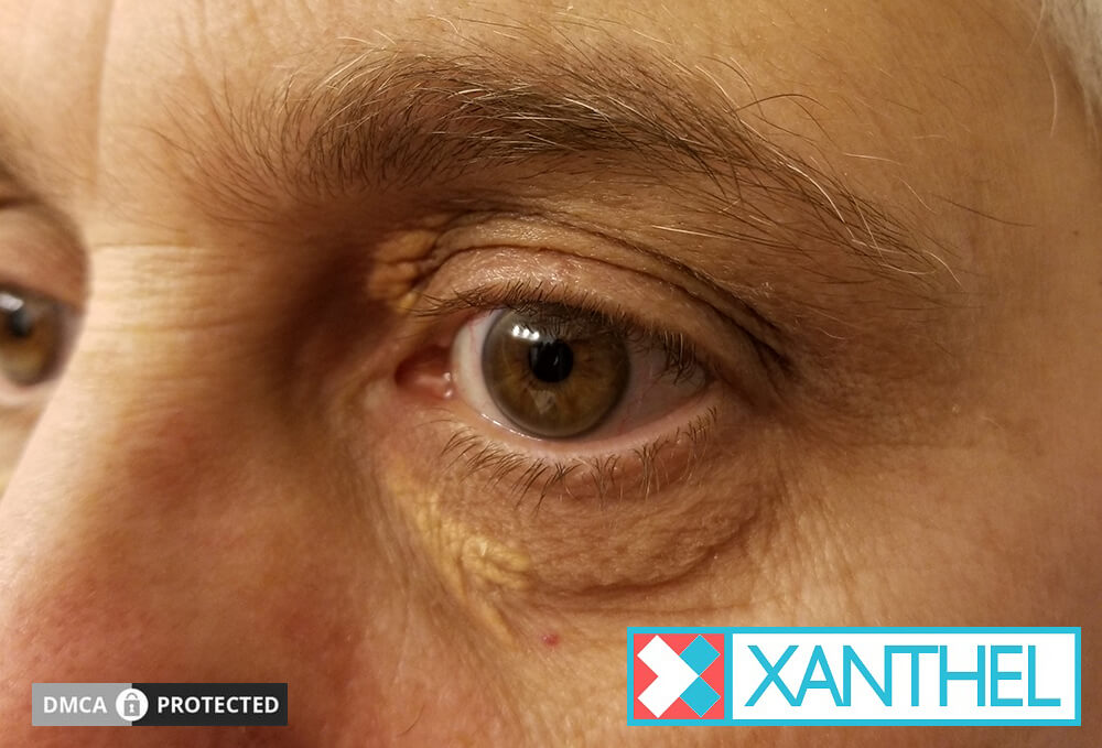
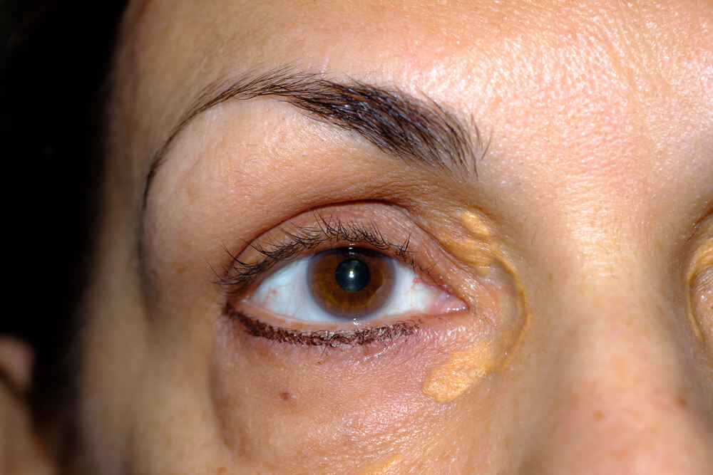
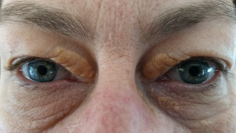
Xanthelasma are soft, yellow, flat or slightly raised plaques on the eyelids, usually near the inner corners and often on both sides. They’re a harmless buildup of fat-filled cells in the skin, and the most common type of xanthoma.
The full medical name is xanthelasma palpebrarum, meaning yellow plaques of the eyelids. People also call them cholesterol deposits on the eyelids, yellow spots on the eyelids or fatty deposits around the eyes. They belong to the xanthoma family, fatty deposits that can form in skin and tendons, and they’re its most common member.[1]
They don’t hurt, itch or affect your vision, and they’re not contagious or cancerous. For most people they’re a cosmetic concern, with one important caveat: they can be a visible sign of a blood fat problem. Our guides to xanthelasma, what’s xanthelasma and xanthelasma facts cover the condition from other angles.
What Are Xanthelasma Made Of?
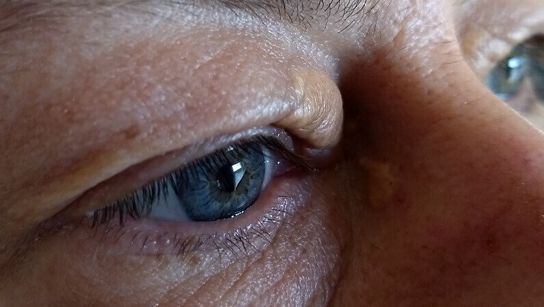
They’re made of foam cells: immune cells called macrophages that have swallowed so much fat they’ve become swollen and foamy. These cells cluster around tiny blood vessels in the deeper layer of the eyelid skin, with some inflammation.
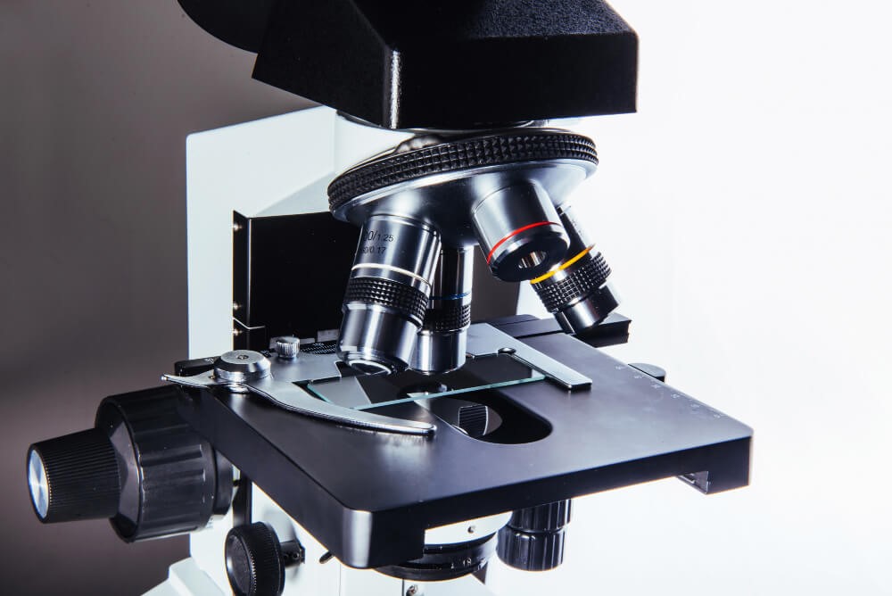
Under the microscope, xanthelasma is a collection of these fat-laden macrophages sitting in the reticular dermis, the deeper part of the skin, gathered around small vessels.[1] The fat they hold includes cholesterol, which is why the plaques are often called cholesterol deposits.
This has two practical consequences. The plaques look yellow because you’re seeing stored fat through very thin skin. And they can’t be squeezed or popped, because the fat is locked inside living cells rather than sitting in a pocket. Our guide to what is xanthelasma gives the short definition.

How Do Xanthelasma Form?
Fat carried in the blood leaks out of tiny eyelid blood vessels, which blinking squeezes and which become leakier with age. Immune cells swallow the escaped fat, become overloaded, and cluster into a yellow plaque.
- Fat travels in the blood. Cholesterol and other fats are carried in particles such as LDL.
- It leaks into the eyelid. Blinking compresses the small vessels, and with age they leak more.[1]
- Immune cells respond. Macrophages swallow the escaped fat.
- They overload. Unable to process it all, they swell into foam cells.
- A plaque appears. The foam cells gather around the vessels, with some inflammation, and the yellow patch forms.[1]
Higher blood fats mean more fat to leak, which is why cholesterol matters. But even normal amounts can slowly escape through leakier vessels, which is why some people get xanthelasma with normal blood tests. Our guide on how xanthelasma forms goes step by step.
Why Do Xanthelasma Appear On The Eyelids?
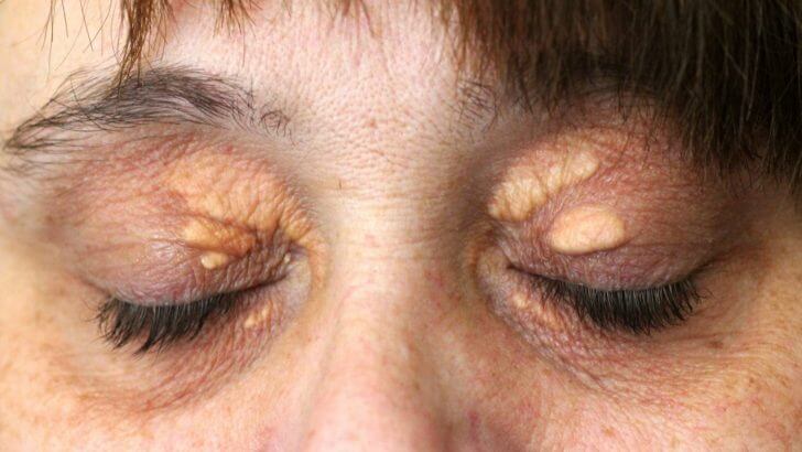
Because eyelid skin is the thinnest on the body and constantly moving. Blinking keeps squeezing its tiny blood vessels, which encourages fat to leak out, and even a thin layer of fat-filled cells shows through as yellow.
The inner corner of the upper lid is the most common spot, and the plaques are usually on both sides. They’re more common on the upper than the lower lid.[4] Our guide to what xanthelasma looks like covers the typical positions and appearances, from faint flat patches to firmer raised plaques.

Who Gets Xanthelasma?
They affect about 1.1% of women and 0.3% of men, so women get them close to four times as often. They usually appear between 35 and 55, though they’re seen from about 20 to 70.
Xanthelasma is seen from about age 20 to 70, most often between 35 and 55.[1] The American Academy of Ophthalmology describes a peak between 30 and 50.[4] When they appear in younger people, doctors look harder for an inherited cholesterol condition.[1]
The old idea that xanthelasma mainly affects particular ethnic groups isn’t something we’ve found good evidence for, so we don’t repeat it. Our guide on whether xanthelasma is common looks at the numbers.

What Do Xanthelasma Mean For Your Health?
About half of people with them have a blood fat disorder, such as raised LDL or low HDL, and they’ve been linked with higher cholesterol and a higher risk of heart disease. The other half have normal blood tests. A check-up settles which group you’re in.

About 50% of people with xanthelasma have a primary or secondary lipid disorder.[1] Reported figures vary, with 25% to 70% of patients having normal blood fats when first seen.[4] Pooling 15 studies of 854 people, researchers found higher total cholesterol, LDL and apolipoprotein B in people with xanthelasma, lower apolipoprotein A1, and thicker carotid artery walls.[2]
A Danish study that followed almost 13,000 people for decades found xanthelasma was linked with a higher risk of heart attack and heart disease, regardless of cholesterol.[3] That’s why a check-up, with a fasting lipid profile, liver, thyroid and blood sugar tests, is worthwhile.[1] Our guide to what xanthelasma indicates explains more, and causes of xanthelasma covers each cause.

Are Xanthelasma A Sign Of Ageing?
Not exactly. They’re more common in midlife, partly because eyelid blood vessels get leakier with age, but they aren’t a normal part of getting older, and many older people never develop them.
Age plays a part through the vessels: researchers think eyelid capillaries become leakier with age, letting fat escape more easily.[1] That helps explain why most people notice xanthelasma between 35 and 55.[1] But blood fats, genes and other health conditions matter too, so xanthelasma is a reason for a check-up, not something to dismiss as ageing.
How Do Xanthelasma Change Over Time?
They usually start small and faint and slowly grow, thicken and spread, sometimes merging or appearing on the other eye. They rarely fade on their own.
Because the fat sits inside cells in the skin, there’s no easy way for the body to clear it, so plaques tend to stay or slowly enlarge. Older plaques are more likely to be firmer and deeper, which matters when it comes to removal. Our guide on how fast xanthelasma grows covers the timeline.
How Are Xanthelasma Different From Other Eyelid Bumps?
Xanthelasma are yellow, soft and usually at the inner corners on both sides. Most other eyelid bumps are white, skin-colored or red, feel firm or tender, or sit in different places.
Points To Xanthelasma
- Yellow to yellow-orange color
- Soft, flat or slightly raised
- Inner corners of the upper lids
- Similar on both sides
Points To Something Else
- Tiny, firm, pure white dots (often milia)
- Small skin-colored clusters on the lower lids (often syringomas)
- A red, tender lump at the lash line (often a stye)
- Hard, bleeding or fast-growing patches
Syringomas are one of the conditions doctors consider alongside xanthelasma, and a few rarer conditions can cause yellow plaques too.[4] A doctor can usually tell by looking, and a small biopsy settles any doubt.[1]

Are Xanthelasma Dangerous?
The plaques themselves aren’t dangerous: they’re benign, not cancer, not contagious and don’t affect vision. What matters is what they may point to, which is why a cholesterol check is worth having.
Rarely, other conditions can cause yellow plaques around the eyes, such as necrobiotic xanthogranuloma, which is why a doctor should confirm the diagnosis.[4] If your plaques are hard, bleeding, growing fast or appearing elsewhere on the body, see a doctor promptly. Our guides on whether xanthelasma is dangerous and whether xanthelasma can be cancerous put the risks in proportion.

How Do You Get Rid Of Xanthelasma?
With a removal method: at a clinic by laser, surgery, chemical peel or radiofrequency, or at home with a cream made for xanthelasma. Lowering cholesterol protects your health but rarely clears existing plaques.
Clinic methods work in the right hands. A review of 49 studies found CO2 laser gave more than 75% improvement in over 90% of patients, with skin color change the most common complication, and surgery used for larger, deeper plaques.[5] Surgery can change the eyelid’s position, and freezing or chemical methods can scar or discolor the skin.[1] Home remedies are a different story: a review of 39 garlic burn cases found mostly second-degree burns, some with scarring.[6]
Our guides to the removal of xanthelasma and what doctor removes xanthelasma compare the clinic options, and xanthelasma cream covers the at-home route.
Once your doctor has checked your cholesterol, if you’d like to deal with your plaques at home, this is the cream we make for xanthelasma.

Ways To Get Rid Of Xanthelasma Compared
| Option | Where | What The Evidence Shows | Main Downsides |
|---|---|---|---|
| Xanthel® removal cream | At home | Cosmetic cream applied only to the plaque; results vary | About one to two weeks of healing; cause needs a doctor’s check |
| CO2 laser | Clinic | Over 75% improvement in over 90% of patients | Skin color change; appointments and cost |
| Surgery | Clinic | Used for larger, deeper plaques | Scarring; eyelid position changes |
| Chemical peel | Clinic | Best on small plaques | Scarring and discoloration |
| Garlic, vinegar, oils | At home | No evidence of removal; burns reported | Burns and scarring |
| Lowering cholesterol | With your doctor | Protects your health | Rarely clears existing plaques |
Do Xanthelasma Come Back After Removal?
They can. Recurrence is generally under 50%, is higher for deeper plaques and after repeated procedures, and has been reported at 13% to 16% after laser.
Recurrence is generally under 50%, and deeper plaques come back more often.[1] Recurrence after laser has been reported at 13% to 16%.[4] Managing any cholesterol, thyroid or blood sugar problem gives you the best chance of fewer new plaques. Our guide on whether xanthelasma comes back covers this in full.

What’s It Like Living With Xanthelasma?
Physically, nothing changes: they don’t hurt or affect vision. But they sit right where people look, and many people find they knock their confidence. Makeup can cover them day to day, and removal means you don’t have to.
A lavender or peach color corrector under a creamy concealer helps neutralize the yellow, especially on flat plaques. Our guide on how to cover xanthelasma goes step by step. Feeling self-conscious about them is perfectly reasonable, and wanting them gone is a good enough reason to remove them once you’ve had your check-up. They’re part of a wider family of fatty deposits; our guide to xanthomas explains how they fit in.

Xanthelasma: What Are They? The Bottom Line
Xanthelasma are harmless yellow plaques of fat-filled cells on the eyelids. They don’t affect your vision, but about half of people with them have a blood fat disorder, so have one check-up, then decide whether to remove them.
They rarely fade on their own, and home remedies can burn the delicate eyelid skin. Xanthel® is a cosmetic cream made for xanthelasma removal at home, sold by Xanthelasma.com. You’ll find it at the foot of this page, or read more about Xanthel® xanthelasma removal cream and browse all our xanthelasma guides.
Common Questions About Xanthelasma
What Are Xanthelasma?
Xanthelasma are soft yellow plaques on the eyelids made of fat-filled immune cells called foam cells. They’re harmless and the most common type of xanthoma.
What Are Xanthelasma Made Of?
They’re made of macrophages, a type of immune cell, that have filled with fat, including cholesterol, and clustered around small blood vessels in the eyelid skin.
Are Xanthelasma Dangerous?
Not in themselves. They’re benign and don’t affect vision, but about half of people with them have a blood fat disorder, so a cholesterol check is worthwhile.
Do Xanthelasma Mean High Cholesterol?
Not always. About half of people with xanthelasma have a blood fat disorder and about half have normal results, so a blood test is the only way to know.
Are Xanthelasma A Sign Of Ageing?
Not exactly. They’re more common in midlife because eyelid vessels get leakier with age, but they aren’t a normal part of ageing and deserve a check-up.
Will Xanthelasma Go Away On Their Own?
Rarely. The fat is locked inside cells in the skin, so plaques usually stay or slowly grow unless they’re removed.
Can Xanthelasma Affect My Eyesight?
No. They sit in the eyelid skin, not the eye, and don’t affect your vision or how your eyelids work.
Can Xanthelasma Be Removed At Home?
Yes. Once your doctor has checked your cholesterol, an at-home cream such as Xanthel®, applied only to the plaque, is one way to remove them.
Sources
- Blair K. Xanthelasma Palpebrarum. StatPearls, National Library of Medicine (NCBI Bookshelf), updated 2026.
- Chang HC, Sung CW, Lin MH. Serum lipids and risk of atherosclerosis in xanthelasma palpebrarum: a systematic review and meta-analysis. Journal of the American Academy of Dermatology, 2020;82(3):596-605.
- Christoffersen M, Frikke-Schmidt R, Schnohr P, Jensen GB, Nordestgaard BG, Tybjaerg-Hansen A. Xanthelasmata, arcus corneae, and ischaemic vascular disease and death in general population: prospective cohort study. BMJ, 2011.
- Patadia A, Rana HS, Stewart K, Yen MT, Lee H. Xanthelasma. EyeWiki, American Academy of Ophthalmology, reviewed 2026.
- Malekzadeh H, Ormseth B, Janis JE. A Practical Review of the Management of Xanthelasma Palpebrarum. Plastic and Reconstructive Surgery Global Open, 2023;11(5):e4982.
- Hitl M, Kladar N, Gavaric N, Srdenovic Conic B, Bozin B. Garlic burn injuries: a systematic review of reported cases. American Journal of Emergency Medicine, 2021.