A Map Of Where It Appears: Inner Corners, Upper And Lower Lids, One Eye Or Both, How Location Shapes Removal, And What Yellow Lumps Elsewhere On The Body Mean
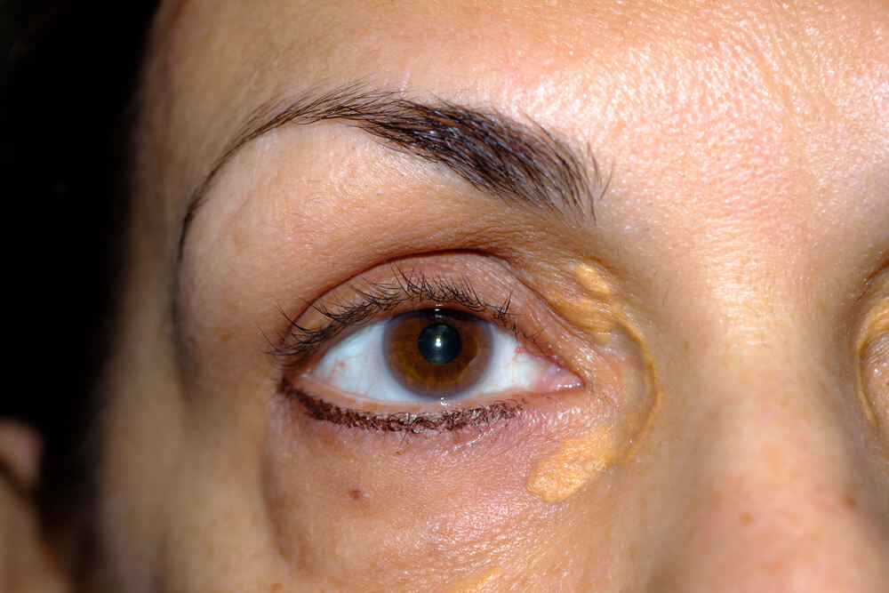
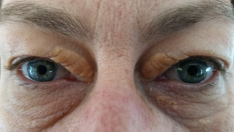
You’ll find xanthelasma on the eyelids, most often on the upper lid near the inner corner by the nose, frequently on both eyes, and sometimes on the lower lids too. It doesn’t appear elsewhere on the body: yellow cholesterol lumps on tendons, elbows, palms or the buttocks are other types of xanthoma, each pointing to a different blood fat problem. Where a plaque sits also affects how it’s best removed.
Xanthelasma appears on the eyelids, most often the upper lid near the inner corner, often on both eyes and sometimes the lower lids. It doesn’t appear on the rest of the body; yellow lumps on tendons, elbows, palms or buttocks are other xanthomas. Plaques near the lid edge usually need an eyelid specialist to remove.
Key Takeaways
- The upper eyelid near the inner corner is the classic site.
- Lower lids can be involved, usually alongside upper-lid plaques.
- Plaques often appear on both eyes.
- Blinking and leaky eyelid vessels help explain why the eyelids are chosen.
- Yellow lumps elsewhere on the body are other xanthomas with different causes.
- Plaques at the lid edge or very near the eye need a specialist to remove.
On This Page 16 sections
Where Can You Find Xanthelasma?
On the eyelids. Most often on the upper lid close to the inner corner, near the nose, and frequently on both sides. Lower lids can be affected too, usually alongside upper-lid plaques.
StatPearls, the National Library of Medicine’s clinical reference, describes xanthelasma as soft, yellowish, flat or slightly raised plaques, usually on the upper lids near the inner corner and sometimes on the lower lids.[1] Its full medical name, xanthelasma palpebrarum, means “yellow plates of the eyelids”, which says exactly where you’ll find it.
Our pages on xanthelasma eyelid and xanthelasma on eyelids cover the eyelid form in depth. This page is the map: where it appears, where it doesn’t, and why location matters.
Why Is Xanthelasma Usually Found At The Inner Corner?
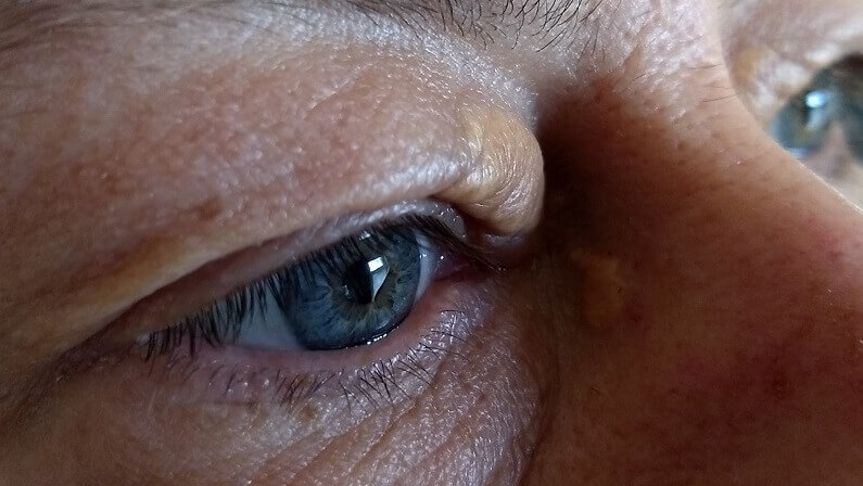
The inner corner of the upper lid is the classic spot, though the exact reason isn’t proven. Constant movement of the lid during blinking and the fine network of vessels there are the leading explanations.
StatPearls describes how blinking compresses the eyelid’s tiny capillaries, which become leakier with age, letting cholesterol-carrying particles into the skin where immune cells swallow them and become foam cells.[1] Plaques typically start as a small flat patch at the inner corner and may widen outward along the lid from there.
Our page on what xanthelasma looks like shows typical inner-corner plaques, and our page on whether xanthelasma can be flat explains early, flat patches.

Is Xanthelasma Found On The Upper Or Lower Eyelid?
Both, but the upper lid far more often. Lower-lid plaques are less common and usually appear alongside upper-lid ones, sometimes forming a ring around the inner corner.
Upper Lid
- The most common site
- Near the inner corner, by the nose
- Can sit in or above the lid fold
- May widen along the lid over time
Lower Lid
- Less common
- Usually with upper-lid plaques
- Near the inner corner or under the eye
- Can be harder to cover with makeup
That pattern follows StatPearls.[1] Our guide to getting rid of xanthelasma under the eyes covers lower-lid plaques in detail.

Is Xanthelasma Found On One Eye Or Both?
Often both, sometimes in a near-mirror pattern. But it can start on one side, and some people only ever have it on one eye.
Plaques frequently appear on both eyes because whatever drives them, a lipid disorder or local factors in the eyelid skin, affects both sides. When they start on one side, a matching plaque often follows on the other over months or years.
A single yellow spot on one lid is still most likely xanthelasma if it’s soft, yellow and painless, but a doctor should confirm it. Our page on eye xanthelasma covers what it means for the eyes themselves.
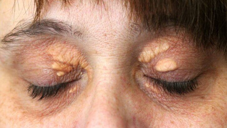
Can Xanthelasma Be Found In The Lid Fold, At The Lash Line Or Near The Eye?
Yes. Plaques can sit in the upper lid crease, extend toward the lash line, or lie very close to the eye at the inner corner. None of these affect your eyesight, but they matter a lot for removal.
- In the lid fold. Partly hidden when the eye is open, more visible when closed.
- Near the lash line. Removal here needs extra care to protect the lid margin.
- At the inner corner, close to the eye. Often best left to an eyelid specialist.
- Effect on sight. None; the plaques sit in the skin, not the eye.[1]
StatPearls warns that removing too much eyelid skin can turn the lid outward or pull it away from the eye, which is why position guides the choice of method.[1]

Does Xanthelasma Spread To New Places?
Only around the eyelids. Plaques can widen along the lid, appear on the other eye or the lower lid, and merge into bands, but xanthelasma doesn’t spread to the cheeks, face or body.

StatPearls describes the plaques as tending to stay the same or slowly progress over time.[1] Flat yellow patches spreading well beyond the eyelids, onto the neck, trunk or skin folds, aren’t typical of xanthelasma and should be checked by a doctor.
Our page on whether xanthelasma spreads looks at the pattern in more detail.

Why Is Xanthelasma Found On The Eyelids?
Most likely because of blinking and leaky vessels, plus very thin skin. Every blink squeezes the lid’s tiny capillaries, they leak more with age, and eyelid skin is thin enough for the yellow foam cells to show through.
That’s the mechanism StatPearls describes.[1] Higher blood cholesterol and lower HDL mean more material to build up, but local factors matter too, since about half of people with xanthelasma have normal blood fats.[1]
Our pages on how xanthelasma forms and what causes cholesterol deposits around the eyes explain the process.

What If You Find Yellow Lumps Elsewhere On Your Body?
Then they’re not xanthelasma but another type of xanthoma, and the location points to the cause. Tendon lumps suggest inherited high cholesterol; palm creases suggest dysbetalipoproteinemia; sudden crops of bumps suggest very high triglycerides.
- Achilles tendons, knuckles, elbows, knees. Tendon xanthomas, a classic sign of familial hypercholesterolemia, which affects about 1 in 250 to 1 in 500 people.[3]
- Palm and finger creases. Palmar xanthomas, linked with dysbetalipoproteinemia; they fade once it’s managed.[4]
- Elbows, knees, buttocks. Tuberous xanthomas, also linked with dysbetalipoproteinemia.[4]
- Buttocks, arms, legs, in crops. Eruptive xanthomas, with triglycerides above 1,000 mg/dL and a pancreatitis risk.[5]
Any of these deserves a prompt doctor’s visit. Our pages on tendon xanthomas, palmar xanthoma, eruptive xanthoma and skin xanthomas explain each.

Where You Find Xanthelasma And Other Xanthomas
| Where | What It Is | Look | Linked With |
|---|---|---|---|
| Eyelids, inner corners | Xanthelasma | Soft, flat or slightly raised yellow plaques | A lipid disorder in about half |
| Achilles tendons, knuckles | Tendon xanthoma | Firm lumps that move with the tendon | Familial hypercholesterolemia |
| Palm and finger creases | Palmar xanthoma | Yellow lines | Dysbetalipoproteinemia |
| Elbows, knees, buttocks | Tuberous xanthoma | Firm yellow nodules | Dysbetalipoproteinemia |
| Buttocks, arms, legs | Eruptive xanthoma | Sudden crops of small yellow-red bumps | Very high triglycerides |
How Do You Check Your Eyelids For Xanthelasma?
In daylight, without makeup, look at both upper and lower lids with your eyes open and then gently closed. Early plaques are flat, pale yellow and easy to miss in warm indoor light.
- Use daylight. Stand by a window; warm bulbs exaggerate yellow and hide edges.
- Remove makeup. Concealer can hide or outline a plaque.
- Look with eyes open. Check the inner corners and the lower lids.
- Close your eyes gently. Plaques in the lid fold show up when the lid is flat.
- Compare both sides. Plaques often appear on both eyes.
- Take photos. Repeat every few months to spot new or growing patches.
If you find a soft, yellow, painless patch, book one visit with your doctor to confirm it and test your blood fats.

What Else Is Found In The Same Places As Xanthelasma?
Milia, syringomas, sebaceous hyperplasia, styes and, rarely, skin cancers can all appear on or around the eyelids. Most look different from soft yellow xanthelasma, and a doctor can usually tell them apart on sight.
- Milia. Tiny, hard, white beads of keratin.
- Syringomas. Small, firm, skin-colored bumps, often under the eyes.
- Sebaceous hyperplasia. Small yellowish bumps, often with a central dip.
- Styes and chalazia. Red, tender or swollen lumps at the lid margin.
- Skin cancers. Rarely, a firm, pearly, bleeding or ulcerated spot on the lid.
StatPearls reserves a biopsy for cases where the diagnosis is uncertain.[1] Our page on what can be mistaken for xanthelasma shows the differences.

Does Where You Find Xanthelasma Say Anything About Your Health?
Not much on its own: an upper-lid or lower-lid plaque carries the same meaning. What matters is that xanthelasma anywhere on the lids is a reason to check your blood fats, and tendon lumps or a young age make that more urgent.
StatPearls reports a primary or secondary lipid disorder in about half of people with xanthelasma and recommends a fasting lipid profile, liver, thyroid and glucose tests.[1] In one clinic study, 54.8% had normal total cholesterol, though HDL was lower than in people without xanthelasma.[2]
If you also find lumps on your tendons, familial hypercholesterolemia is likely and early care makes a real difference.[3] Our page on whether xanthelasma indicates raised cholesterol explains the results.

How Does Where Xanthelasma Is Found Affect Removal?
Plaques on the open eyelid skin, away from the lash line, have the most options: laser, peels, freezing or a cream made for xanthelasma removal at home. Plaques at the lid edge, very near the eye or large enough to need cutting out are best with an eyelid specialist.
A 2023 review of 49 studies found CO2 laser gave more than 75% improvement in over 90% of patients, with surgery recommended for larger, deeper plaques; a change in skin color was the most common complication.[6] Plaques return after removal in generally fewer than half of cases.[1]

Our pages on how xanthelasma is removed, removing it without surgery and our xanthelasma removal cream guide help you choose.

If your doctor has checked the cause and your plaques sit on the eyelid skin away from the lash line, this is the cream we make for removing them at home.
When Should You See A Doctor About Where You Found Xanthelasma?
Once for any new eyelid plaque, for the diagnosis and blood tests. Go promptly if you find yellow lumps on your tendons, palms or body, if spots appeared suddenly or in crops, or if a mark is firm, bleeding or changing fast.
- Tendon lumps or young age. Possible inherited high cholesterol.[3]
- Sudden crops of yellow bumps. Possible very high triglycerides.[5]
- Patches spreading beyond the lids. Not typical of xanthelasma; get them checked.
- Unusual eyelid marks. Firm, pearly, bleeding or ulcerated spots need a closer look.

What’s The Bottom Line On Where You Can Find Xanthelasma?
You’ll find xanthelasma on the eyelids, most often the upper lid near the inner corner, frequently on both eyes and sometimes the lower lids. It spreads only around the lids. Yellow lumps on tendons, palms, elbows or the buttocks are other xanthomas with their own causes. Wherever it sits, get your blood fats checked, and let position guide removal: open lid skin has the most options, the lid edge needs a specialist.
Xanthel® is a cosmetic cream made for xanthelasma removal at home, sold by Xanthelasma.com. You’ll find it in the removal section above and at the foot of this page, or read more about Xanthel® for xanthelasma on the eyelids. For everything else, see our complete xanthelasma guide, whether xanthelasma can disappear, or browse all our xanthelasma guides.
Common Questions About Where You Can Find Xanthelasma
Where Does Xanthelasma Usually Appear?
On the upper eyelid near the inner corner, often on both eyes, and sometimes on the lower lids.
Why Does Xanthelasma Appear On The Eyelids?
Blinking squeezes the lid’s tiny vessels, which leak more with age, and eyelid skin is thin enough for the yellow to show.
Can Xanthelasma Appear On The Lower Eyelid?
Yes, though less often than the upper lid, and usually alongside upper-lid plaques.
Does Xanthelasma Appear On Both Eyes?
Often, sometimes in a mirror pattern, though it can start on one side or stay on one eye.
Can Xanthelasma Appear Anywhere Other Than The Eyelids?
No; yellow cholesterol lumps elsewhere on the body are other types of xanthoma with different causes.
Does Where Xanthelasma Is Affect Your Eyesight?
No, the plaques sit in the eyelid skin, not the eye, wherever they are on the lid.
Does Location Change How Xanthelasma Is Removed?
Yes; plaques on open lid skin have the most options, while those at the lid edge or near the eye need an eyelid specialist.
What Is A Yellow Spot Near The Eye That Isn’t Typical?
It could be milia, syringomas, sebaceous hyperplasia or, rarely, a skin cancer, so have a doctor look at it.
Sources
- Blair K. Xanthelasma Palpebrarum. StatPearls, National Library of Medicine (NCBI Bookshelf), updated 2026.
- Kavoussi H, Ebrahimi A, Rezaei M, Ramezani M, Najafi B, Kavoussi R. Serum lipid profile and clinical characteristics of patients with xanthelasma palpebrarum. Anais Brasileiros de Dermatologia, 2016;91(4):468-471.
- Ison HE, Clarke SL, Knowles JW. Familial Hypercholesterolemia. GeneReviews, National Library of Medicine (NCBI Bookshelf), revised 2025.
- Javvaji A, Can AS, Sharma S. Dysbetalipoproteinemia. StatPearls, National Library of Medicine (NCBI Bookshelf), updated 2024.
- Mishra C, Tripathy K. Lipemia Retinalis. StatPearls, National Library of Medicine (NCBI Bookshelf), updated 2026.
- Malekzadeh H, Ormseth B, Janis JE. A Practical Review of the Management of Xanthelasma Palpebrarum. Plastic and Reconstructive Surgery Global Open, 2023;11(5):e4982.