Flat, Raised Or Lumpy: How To Recognize Each Shape, What It Means, And Why Thickness Matters For Removal
Yes, xanthelasma can be flat. Many plaques start as soft, flat yellow patches on the eyelids that sit level with the skin, while others are slightly raised or form firmer lumps. Flat xanthelasma is still xanthelasma, with the same health links, but its shape and thickness do make a difference when you want to get rid of it.
Yes. Xanthelasma can be completely flat, slightly raised or nodular. Flat plaques are soft yellow patches, usually near the inner corner of the upper eyelids. Shape doesn’t change what xanthelasma means for your health, but thin, flat plaques are generally the easiest to remove, and thicker ones are more likely to need surgery and come back.
Key Takeaways
- Xanthelasma can be flat and soft, slightly raised, or firm and nodular.
- Flat plaques are usually yellow, painless and near the inner corner of the eyelid.
- A flat plaque means the same for your health as a raised one.
- Plaques can thicken, spread and merge over time.
- Thin, superficial plaques suit more removal methods than thick ones.
- Deeper plaques are more likely to come back after removal.
On This Page 14 sections
Can Xanthelasma Be Flat?
Yes. Xanthelasma can be flat and soft, sitting level with the surrounding skin, or it can be raised, firm and nodular. Doctors describe both forms, and a flat yellow patch on the eyelid is a very typical way for xanthelasma to look.
Xanthelasma palpebrarum is a buildup of fat-filled immune cells in the eyelid skin. It shows up as yellowish patches or plaques, and medical reviews describe it as ranging from flat and soft to nodular and semi-solid.[2] Many people first notice a faint, flat yellow mark that’s easy to mistake for a shadow or a skin tone change, and only later see it thicken or spread.
It’s common enough that plenty of people have it without knowing what it is. It affects around 1.1% of women and 0.3% of men,[1] and it can start anywhere from the teens to the seventies, most often between 30 and 50.[2] Our main guide to xanthelasma covers the condition as a whole, and is xanthelasma common looks at who gets it.
So if you’ve spotted soft, flat yellow patches near the inner corners of your eyes, xanthelasma is the most likely explanation. A doctor can usually confirm it simply by looking.[1]

What Does Flat Xanthelasma Look Like?
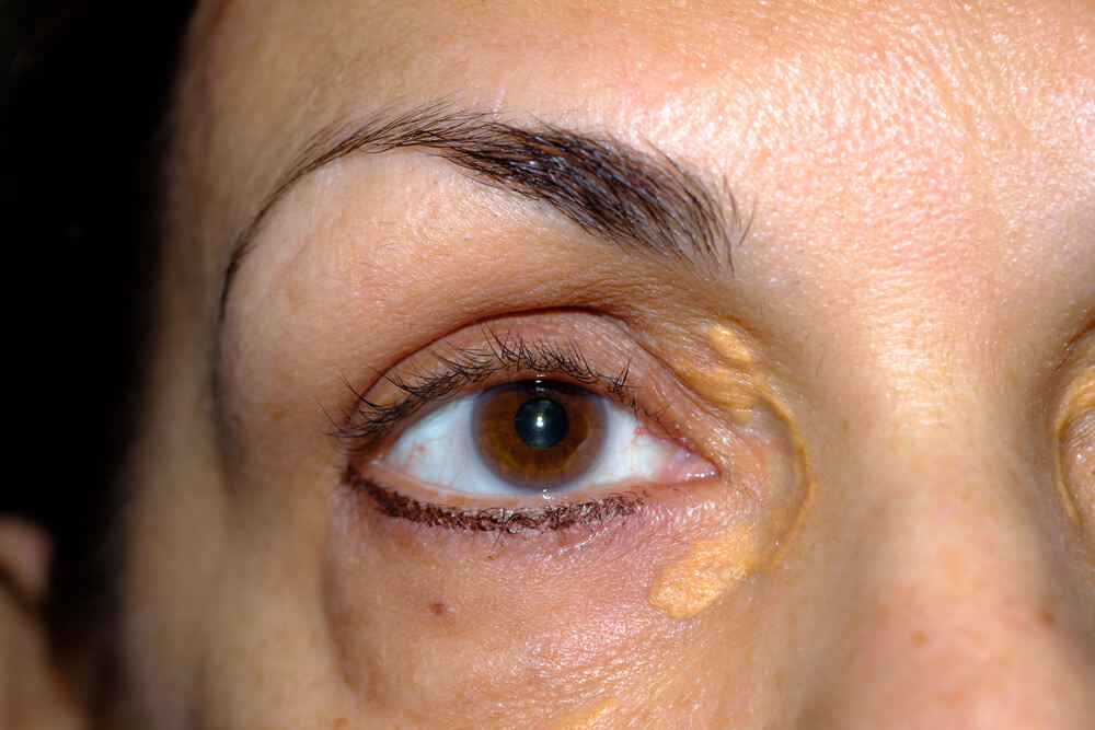
Flat xanthelasma looks like a soft, yellow or yellow-orange patch with fairly clear edges, usually on the upper eyelid near the inner corner and often on both sides. It doesn’t hurt, itch or bleed.
The features that point to flat xanthelasma are:
- Color. Pale yellow to yellow-orange, like a stain in the skin, because you’re seeing stored fat through very thin skin.
- Place. Most often the inner corner of the upper eyelid, sometimes the lower lid too.[2]
- Symmetry. It usually appears on both eyes in a similar position.[2]
- Feel. Soft and level with the skin, or barely raised, with no hard lump underneath.
- Comfort. No pain, itching, crusting or bleeding.
Flat plaques are easiest to see in good daylight and can be hard to spot in photos. Many people notice them first when applying makeup or looking closely in a mirror. Our guides on what xanthelasma looks like and xanthelasma symptoms describe the signs in more detail.

What Shapes Can Xanthelasma Take?
Xanthelasma ranges from flat marks (macules) through soft plaques to raised bumps (papules) and firm lumps (nodules). These are points on the same scale: the more fat-filled cells collect and the deeper they reach, the thicker the plaque.
Dermatologists describe xanthelasma by its shape and depth:
- Macular (flat). Level with the skin, soft and thin. Often the earliest stage.
- Flat plaque. A broader, soft yellow area that may be very slightly raised.
- Papular. Small, clearly raised yellow bumps.
- Nodular. Firmer, thicker lumps that can feel semi-solid.[2]
Why does depth vary? The fat-filled foam cells gather in the deeper layer of the skin around small blood vessels.[1] As more cells build up, the plaque thickens and can push up above the surface. In some people, the deposit reaches down into the muscle that closes the eyelid: in one surgical series of 95 patients, about a quarter had muscle involvement.[3]
A single person can have more than one shape, for example a flat patch on one side and a raised plaque on the other.

Flat And Raised Xanthelasma Compared
| Shape | How It Looks And Feels | Depth | What It Means For Removal |
|---|---|---|---|
| Flat (macular) | Level with the skin, soft, pale yellow | Usually superficial | Suits the widest range of methods, including at-home removal |
| Flat plaque | Broader soft patch, barely raised | Usually superficial | Most methods suitable; larger areas may need more than one session |
| Papular | Small raised yellow bumps | Moderate | Stronger approaches often needed |
| Nodular | Firm, thick, semi-solid lumps | Deep, sometimes into muscle | Surgery often preferred; higher chance of coming back |
How Do Doctors Grade Xanthelasma?
Doctors often grade xanthelasma from I to IV by how much of the eyelid it covers, and they also note its height, because both affect which removal method suits it.
A widely used grading system describes four stages by location:[2][3]
- Grade I. Plaques on the upper eyelids only.
- Grade II. Plaques that extend into the inner corner of the eye.
- Grade III. Plaques on the inner side of both the upper and lower eyelids.
- Grade IV. Widespread plaques on the inner and outer sides of the upper and lower eyelids.
In the surgical series of 95 patients, 35% were grade I, 35% grade II, 25% grade III and 5% grade IV.[3] Doctors also record how tall the plaque is, since a flat plaque and a raised one in the same spot can need quite different approaches.[2] A flat, grade I plaque is about as small and simple as xanthelasma gets.

Does Flat Xanthelasma Become Raised Over Time?

It can. Xanthelasma tends to stay or grow slowly rather than fade, and over months or years a flat patch can thicken, spread and merge with nearby plaques. Plaques present for longer tend to be firmer and deeper.
Not every flat plaque changes. Some stay small and flat for years. But because the fat-filled cells don’t clear by themselves, many plaques slowly enlarge, and new ones can appear beside them. Removal guides draw a line between soft, recent plaques that have been there under a year, and harder, older plaques that have been there longer, because the older ones are more likely to have reached deeper into the skin.[2]
That’s why many people choose to deal with plaques while they’re still flat. Our guides on whether xanthelasma can get bigger, how fast xanthelasma grows and whether xanthelasma spreads look at how plaques change.

Is Flat Xanthelasma Less Serious Than Raised Xanthelasma?
No. Shape describes how the plaque looks, not what it means. Flat or raised, the plaque is harmless in itself, but about half of people with xanthelasma have a blood fat disorder, so it’s worth a cholesterol check either way.
About 50% of people with xanthelasma have a primary or secondary lipid disorder, such as raised LDL cholesterol or low HDL.[1] In the surgical series, 52% had an abnormal lipid profile.[3] Other conditions such as diabetes, an underactive thyroid and some liver conditions can play a part too.[1]
There’s also a heart link. A Danish study that followed almost 13,000 people for decades found that people with xanthelasma had a higher risk of heart attack and heart disease, regardless of their cholesterol levels.[5] Nothing suggests flat plaques carry less of that link than raised ones, so a flat patch deserves the same simple check: a fasting lipid profile, plus thyroid, liver and blood sugar tests.[1]
The plaques themselves don’t turn into anything harmful. Our guides on whether xanthelasma is dangerous and whether xanthelasma indicates raised cholesterol put the risks in proportion.
What Else Can Cause Flat Yellow Patches Around The Eyes?
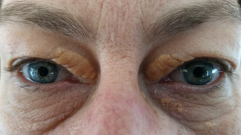
Most flat yellow patches on the inner eyelids are xanthelasma, but a few other conditions can look similar. Patches that are hard, ulcerated, spreading fast, or appearing elsewhere on the body should always be checked by a doctor.
Because flat xanthelasma is subtle, it’s sometimes confused with other things, and other things are sometimes confused with it. A few signs suggest a doctor’s opinion is needed before anything else:
Typical Of Xanthelasma
- Soft yellow patches on the inner eyelids
- On both sides, in a similar spot
- Painless and slow to change
- Smooth surface, no crusting
Worth A Doctor’s Check
- Firm, hard or ulcerated patches
- Rapid growth or bleeding
- Flat yellow patches on the neck, trunk or skin folds
- White rather than yellow spots
Flat yellow patches that spread beyond the eyelids can be a different condition called plane xanthoma, which needs its own medical workup; see plane xanthoma. Our guides to what can look like xanthelasma and whether xanthelasma can be white cover the common lookalikes. If you’re unsure, a biopsy can settle it, though it’s rarely needed.[1]

Why Does Flat Xanthelasma Appear?
Flat xanthelasma forms the same way as raised xanthelasma: fats leak from tiny eyelid blood vessels and are taken up by immune cells, which become fat-filled foam cells. It’s simply an earlier or thinner deposit.
Researchers think the constant pressure of blinking squeezes the small blood vessels in the eyelid, and that these vessels get leakier with age. Fat-carrying particles escape into the skin, macrophages swallow them, and once overloaded they become foam cells clustered around the vessels.[1] When only a thin layer of these cells has built up, the result is a flat yellow patch.
What drives the leak varies from person to person: a blood fat disorder in about half, diabetes, thyroid or liver problems in some, inherited cholesterol conditions in others, especially younger people, and local eyelid factors in people whose blood tests are normal.[1] Our guide to the causes of xanthelasma goes through each one.
How Do You Get Rid Of Flat Xanthelasma?
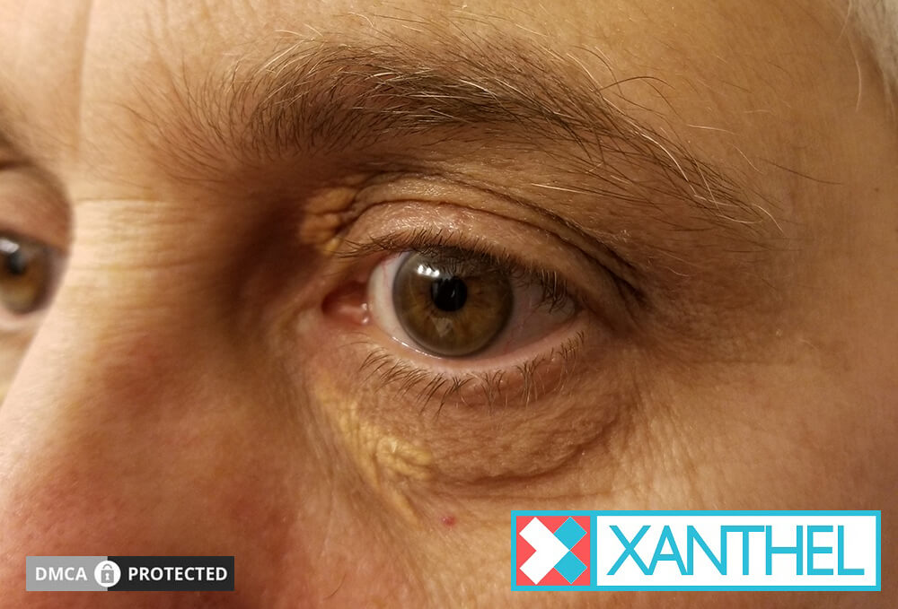
Flat, superficial xanthelasma suits the widest range of removal methods, including laser, freezing, chemical peels and at-home removal. Thick, deep or long-standing plaques are more likely to need surgery.
Thickness is one of the main things a clinician weighs. Guidance from published reviews suggests:[2][3]
- Superficial, soft plaques under 5 mm high, present under a year: laser, radiofrequency, chemical peels or freezing, chosen to suit the person.
- Deep, hard plaques 5 mm or higher, present over a year, or reaching the muscle: surgical removal is usually the better choice.
Flat plaques also need gentler approaches. With chemical peels, for example, a 50% acid strength was enough for flat macular plaques, while raised, nodular ones did best with the strongest concentration.[2] Laser is effective on the right plaques: a review of 49 studies found CO2 laser gave more than 75% improvement in over 90% of patients, with skin color changes the most common complication.[4] Laser is best kept to small, shallow plaques, and larger plaques removed by laser showed more scarring, discoloration and recurrence in one series.[3] Clinic costs vary; see how much xanthelasma removal costs.

Many people with flat plaques would rather not go to a clinic at all. Our guides to the removal of xanthelasma and how to get rid of xanthelasma compare every option, and xanthelasma cream covers the at-home route.
If your doctor has checked your cholesterol and you’d like to deal with flat plaques at home before they thicken, this is the cream we make for xanthelasma.

Will Flat Xanthelasma Go Away On Its Own?
Rarely. Flat plaques don’t usually fade by themselves, and lowering cholesterol tends to protect your health more than it shrinks the plaques. Most stay the same or slowly grow.
The fat in xanthelasma is stored inside living cells in the skin, not floating loose, so there’s no easy way for the body to clear it. Our experience is that established plaques, flat or raised, rarely shrink noticeably with diet or cholesterol-lowering medicine alone. Managing your blood fats is still worth doing for your heart, and it may help limit new plaques, but it usually isn’t a way to remove the ones you have.
That’s why the usual plan has two parts: a check-up for the cause, and a removal method for the plaques if they bother you. Our guide on whether xanthelasma can go away on its own explains more.

Does Flat Xanthelasma Come Back After Removal?
It can, as with any xanthelasma, but deeper plaques come back more often than shallow ones. Recurrence is generally reported at under 50%, and it’s higher after repeated procedures and when all four eyelids are involved.
The figures depend on the plaque and the method. Recurrence after removal is generally under 50%, and deeper plaques return more often.[1] After surgery, one widely quoted report found recurrence of 40% after a first excision, 60% after a second, and 80% when all four eyelids were affected, with about a quarter of recurrences in the first year.[2] Incomplete removal raises the risk.[3]
Because flat plaques are usually shallow and limited, they sit at the better end of that range. Keeping on top of any cholesterol, thyroid or blood sugar problem helps too. Our guide on whether xanthelasma comes back covers recurrence in full.

What’s The Bottom Line On Flat Xanthelasma?
Xanthelasma can be flat, and flat plaques are still xanthelasma, with the same health links as raised ones. They’re usually the thinnest and most superficial plaques, which makes them the simplest to remove.
Have one check-up for cholesterol, thyroid and blood sugar, because that’s about your health. Then, if the flat patches bother you, deal with them before they thicken or spread. Xanthel® is a cosmetic cream made for xanthelasma removal at home, sold by Xanthelasma.com. You’ll find it at the foot of this page, or read more about Xanthel® xanthelasma removal cream and browse all our xanthelasma guides.
Common Questions About Flat Xanthelasma
Can Xanthelasma Be Completely Flat?
Yes. Xanthelasma can sit completely level with the skin as a soft yellow patch, or it can be slightly raised or nodular. Flat plaques are a very typical form.
Is Flat Xanthelasma Less Serious?
No. Shape describes how it looks, not what it means. Flat or raised, the plaque is harmless itself, but about half of people with xanthelasma have a blood fat disorder worth checking.
How Do I Know If A Flat Yellow Patch Is Xanthelasma?
Look for a soft, painless yellow patch near the inner corner of the eyelids, often on both sides. A doctor can usually confirm it just by looking.
Can Flat Xanthelasma Turn Into Raised Xanthelasma?
It can. Plaques often grow slowly, thicken and merge over months or years, and older plaques tend to be firmer and deeper than recent ones.
Is Flat Xanthelasma Easier To Remove?

Generally, yes. Thin, superficial plaques suit more removal methods and tend to come back less often than thick, deep plaques, which are more likely to need surgery.
Does Flat Xanthelasma Mean High Cholesterol?
Not necessarily. About half of people with xanthelasma have a blood fat disorder and about half have normal results, so a simple blood test is the way to find out.
Will Flat Xanthelasma Fade On Its Own?
Rarely. Flat plaques usually stay or slowly grow, and lowering cholesterol tends to protect your heart rather than clear the plaques you already have.
Can I Remove Flat Xanthelasma At Home?
Yes, many people do. Once a doctor has checked your cholesterol, an at-home cream such as Xanthel® is one way to deal with flat plaques before they thicken.
Sources
- Blair K. Xanthelasma Palpebrarum. StatPearls, National Library of Medicine (NCBI Bookshelf), updated 2026.
- Nair PA, Singhal R. Xanthelasma palpebrarum: a brief review. Clinical, Cosmetic and Investigational Dermatology, 2017;11:1-5.
- Outcomes of surgical management of xanthelasma palpebrarum. Archives of Plastic Surgery, 2013;40(4):380-386.
- Malekzadeh H, Ormseth B, Janis JE. A Practical Review of the Management of Xanthelasma Palpebrarum. Plastic and Reconstructive Surgery Global Open, 2023;11(5):e4982.
- Christoffersen M, Frikke-Schmidt R, Schnohr P, Jensen GB, Nordestgaard BG, Tybjaerg-Hansen A. Xanthelasmata, arcus corneae, and ischaemic vascular disease and death in general population: prospective cohort study. BMJ, 2011.